

Colon tumor


Case report
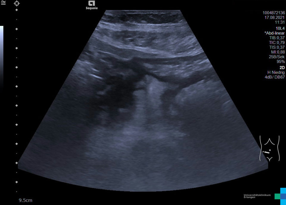
60-year-old female patient with CVID immunoglobulin deficiency. With an unclear increase in inflammatory parameters, multiple liver foci were detected on SPECT-CT. Abdominal sonography revealed solid hepatic lesions. In case of suspected liver metastases, abdominal sonography and intestinal sonography were extended. In the left lower abdomen (video and image), a circumscribed wall thickening with lumen narrowing in the area of the sigmoid colon was noted. Summarizing sonographic findings: Hepatic metastatic sigmoid carcinoma.


