
Venous thrombosis in the leg
-
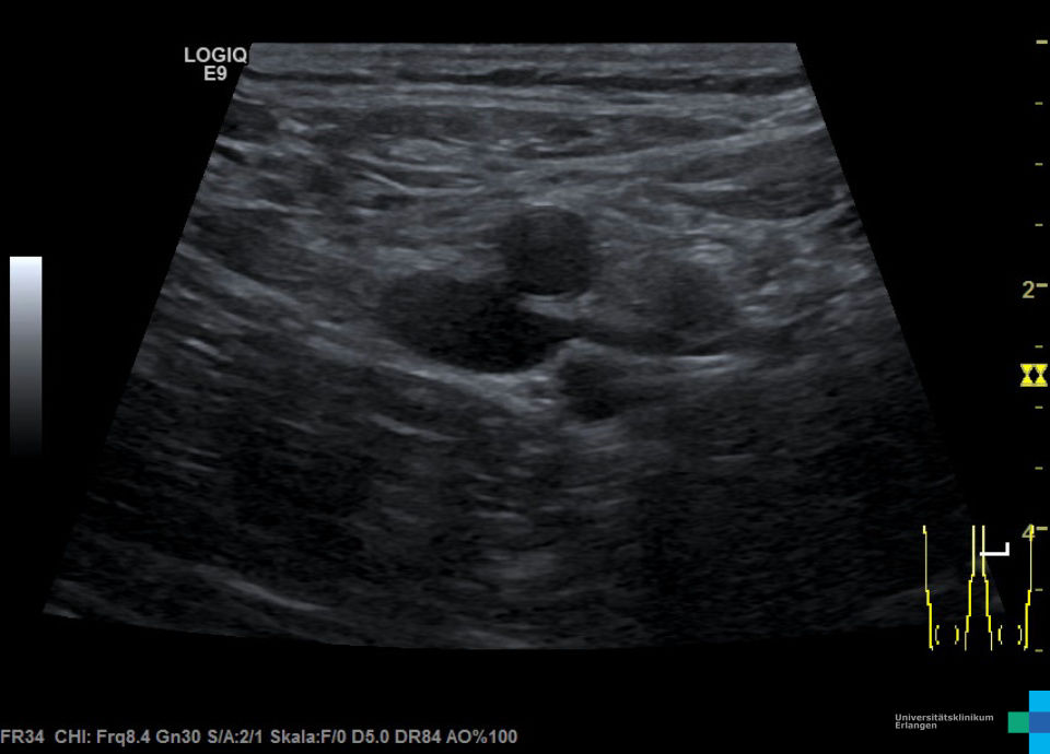
 Freshly occluded thrombosis
 -
 Freshly occluded thrombosis
 -
 Freshly occluded thrombosis (video)
 -
 Long thrombosis (video)
 -
 Fibular vein thrombosis (video)
 -
 Level III thrombus in common femoral vein
 -
 Level III thrombus in common femoral vein
 -
 Thrombophlebitis of the V. saphena parva (video)
 -
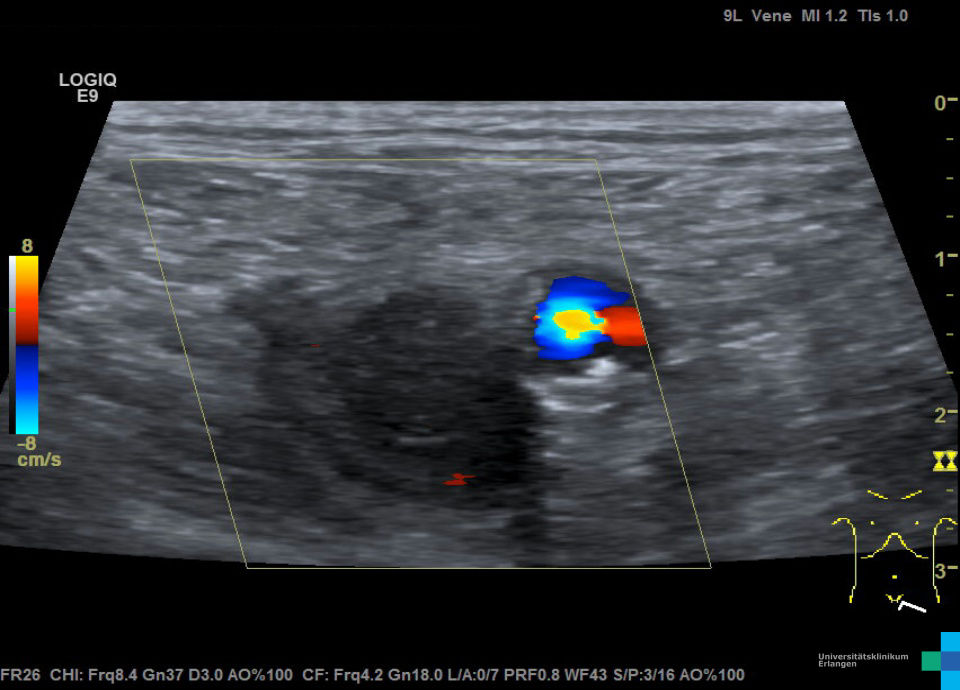
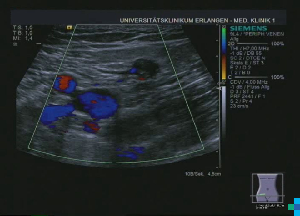
 Thrombosis of the popliteal vein (color doppler video)
 -
 V. poplitea thrombosis (video)
 -
 Thrombosis V. femoralis superficialis right (video)
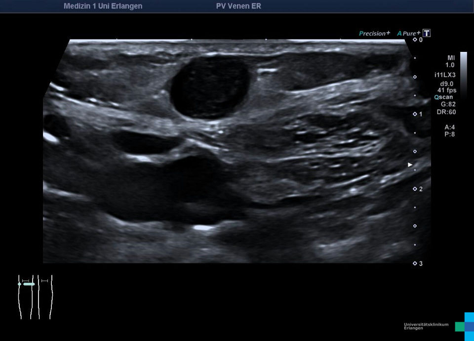
Frisch okkludierende Thrombose der V. femoralis communis (hochfrequenter Linearschallkopf)
Frisch okkludierende Thrombose der V. femoralis communis (hochfrequenter Linearschallkopf)
Frisch okkludierende Thrombose der V. femoralis communis (hochfrequenter Linearschallkopf)
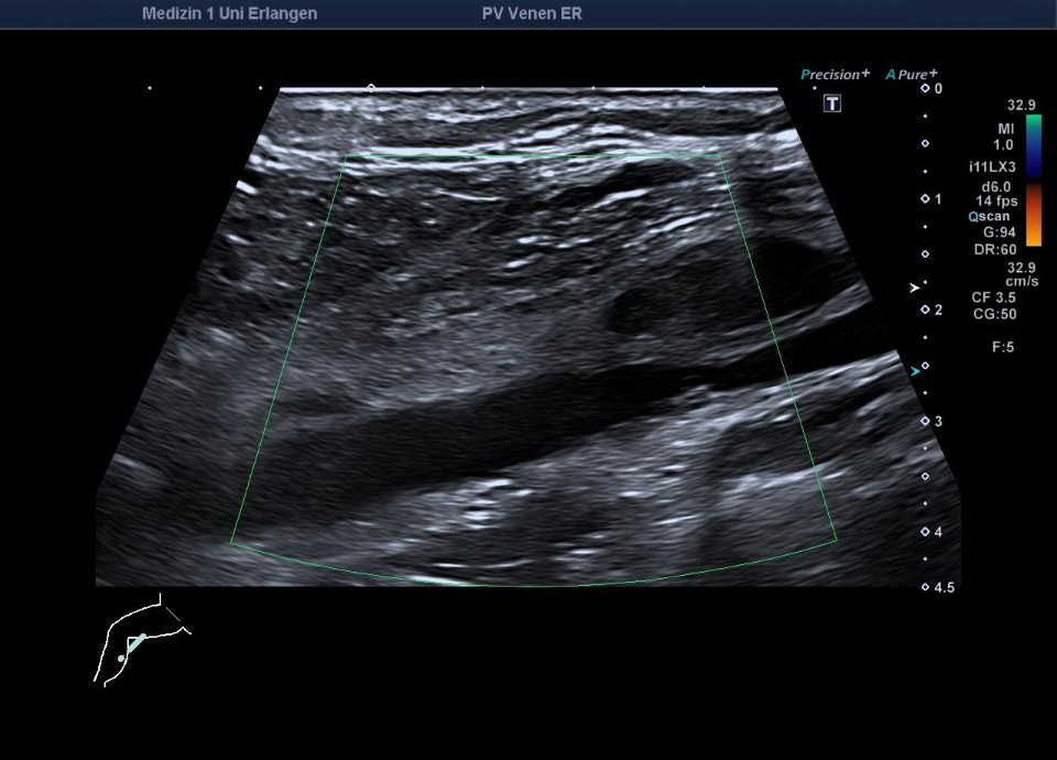
Langstreckige Thrombose der V. saphena magna rechts bis zum Mündungsbereich (hochfrequenter Linearschallkopf)
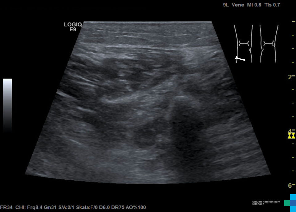
Thrombose der an der Fibula entlang ziehenden V. fibularis (echoarmes, nicht komprimierbares Material in der V. fibularis; hochfrequenter Linearschallkopf)
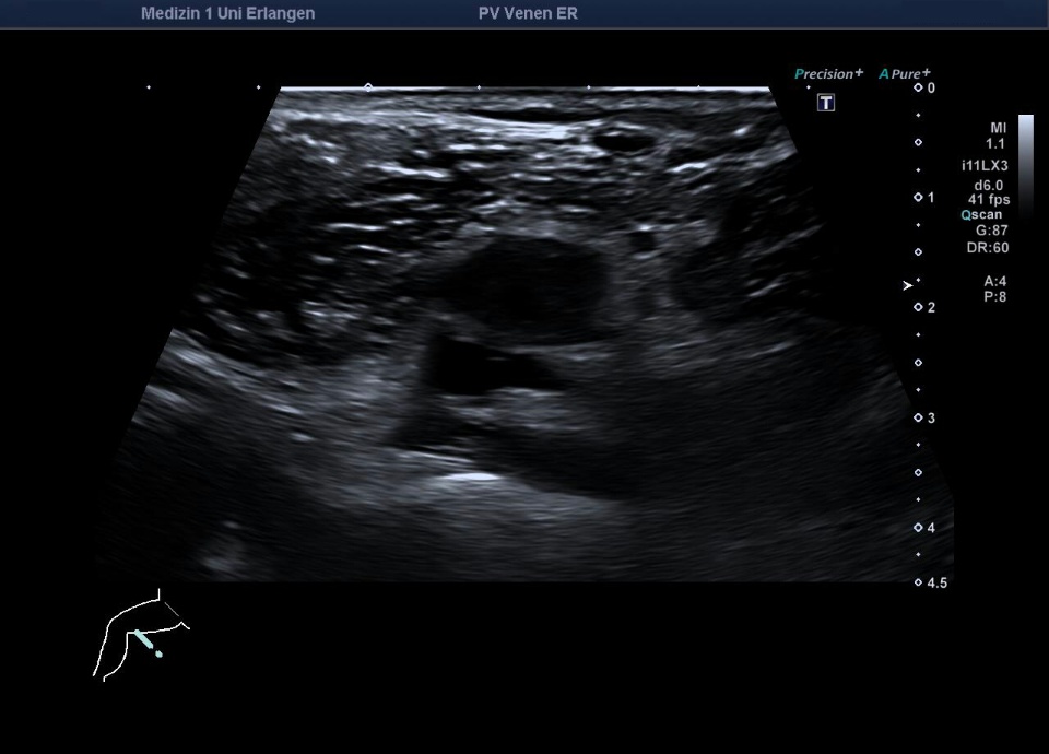
Im thrombosierten Bereich der V. femoralis communis keine Kompression möglich (hochfrequenter Linearschallkopf), siehe Folgebild
Thrombusspitze in der V. femoralis communis in Höhe des Confluens im Längsschnitt (hochfrequenter Linearschallkopf)
Ultraschall, Ultraschallatlas, Ultraschallbilder, Ultraschallvideos, Sonographie, Sonographieatlas, Sonographiebilder, Sonographievideos, Kontrastmittelultraschall, Kontrastmittelsonographie, Medizinische Klinik 1, Uniklinik, Universitätsklinikum, Erlangen