

E. multilocularis
Case 1:
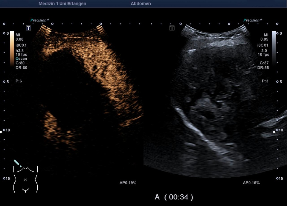
81 year old female patient, increased fatigue and decreased performance since 1 year. CRP, G-GT, Alkal. Phophatase slightly elevated => external MRI with suspicion of malignant liver lesion. Inhomogeneous hepatic cavity on ultrasound => presentation for CEUS. CEUS revealed uncontrasted hepatic mass. In combination with the 2d-image suspicious for echinococcus multilocularis. Patient confirmed 40-years of dog contact. Echinococcus titer positive (IHAT 1:1280). Albendazole therapy started. Patient was not operable due to age and comorbidities.


Case 2:
55-year-old female patient in an external hospital, admitted due to painless jaundice. Bile duct stent was inserted. Slight laboratory increase in gamma-GT, alkaline phosphatase and CRP. Positive echinococcus serology (1:320). B-scan characteristic findings of an echinococcus multilocularis in the liver segments VII / VIII/V/ IVb: hyperechoic, inhomogeneous, irregularly limited mass with small calcifications and cysts. Only the cystic areas are demarcated in CEUS.


