

Diverticulitis
-
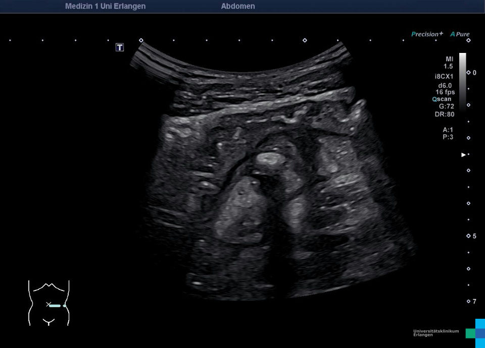
 Diverticulitis of the sigmoid colon
 -
 Diverticulitis with concurring response of the sigma (video)
 -
 Diverticulitis with concurring response of the sigma (video)
 -
 Diverticulitis of the sigmoid colon (video)
 -
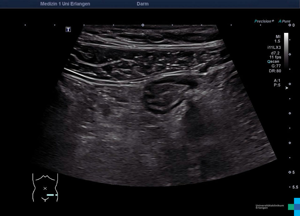
 Acute diverticulitis of the sigmoid colon
 -
 Diverticulitis with abscess
 -
 Paracolic macroabscess (video)
 -
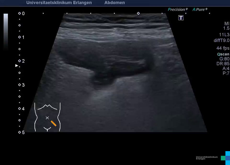
 Complicated sigmoid diverticulitis (type 2a) (video)
 -
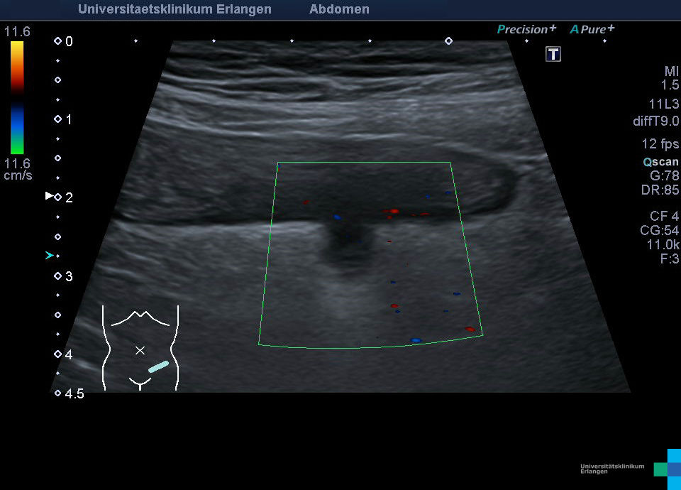
 Complicated sigmoid diverticulitis (type 2a) - color doppler mode
 -
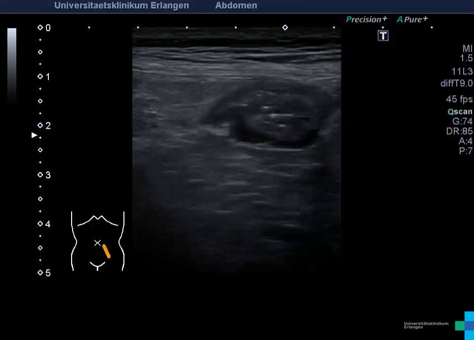
 Diverticulitis of the sigmoid colon (video)
 -
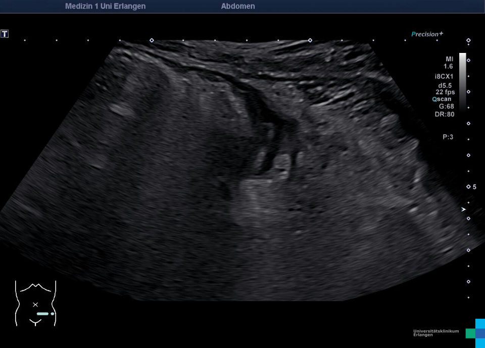
 Diverticulitis of the sigmoid colon
 -
 Acute complicated diverticulitis (abscess)
 -
 Complicated diverticulitis (abscess) (Video)
 -
 Acute diverticulitis
 -
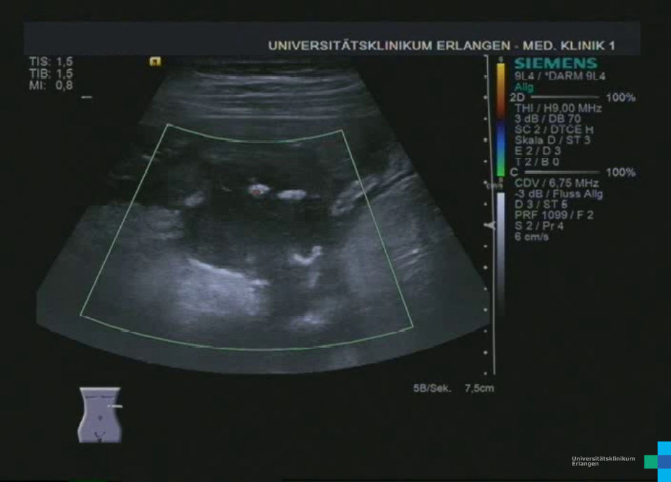
 Acute diverticulitis (color doppler)
 -
 Sigmoid diverticulitis with microabscess (video)
 -
 Sigmoid diverticulitis with microabscess
Sigmadivertikulitis (hochfrequenter Linearschallkopf)
Divertikulitis mit entzündlicher Mitreaktion des Sigmas (hochfrequenter Linearschallkopf)
Divertikulitis mit entzündlicher Mitreaktion des Sigmas und entzündlicher Fettgewebsreaktion (hochfrequenter Linearschallkopf)
Sigmadivertikulitis (hochfrequenter Linearschallkopf)
Akute Divertikulitis mit echoreicher Entzündungsreaktion des mesenterialen Fettgewebes (hochfrequenter Linearschallkopf)
Divertikulitis mit Abszess (hochfrequenter Linearschallkopf)
Paracolischer Makroabszess mit entzündlicher Mitreaktion des Kolons bei zunächst vermuteter Divertikulitis im Bereich der linken Flexur (hochfrequenter Linearschallkopf)
Unkomplizierte Sigmadivertikulitis mit mesenterialer Entzündungsreaktion und Koprolith im Divertikel (Farbdoppler im nächsten Bild)
Unkomplizierte Sigmadivertikulitis mit mesenterialer Entzündungsreaktion (Hyperämie im Farb-Doppler)
Ultraschall, Ultraschallatlas, Ultraschallbilder, Ultraschallvideos, Sonographie, Sonographieatlas, Sonographiebilder, Sonographievideos, Kontrastmittelultraschall, Kontrastmittelsonographie, Medizinische Klinik 1, Uniklinik, Universitätsklinikum, Erlangen