
Budd-Chiari syndrome
Case 1:


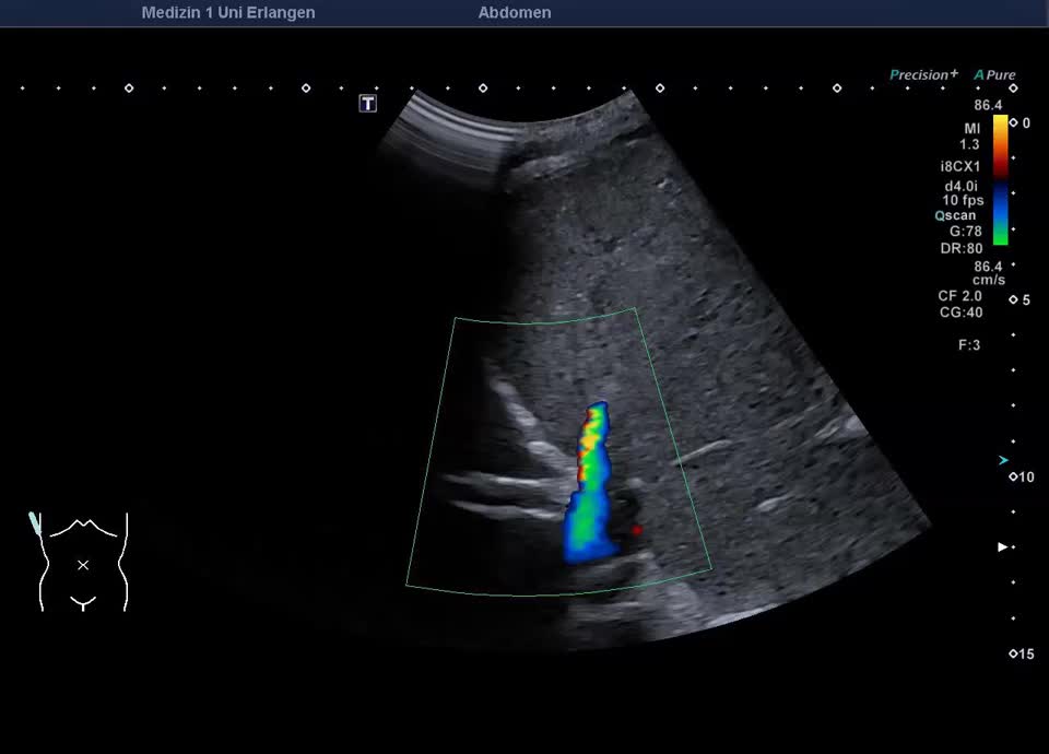
Case 2:
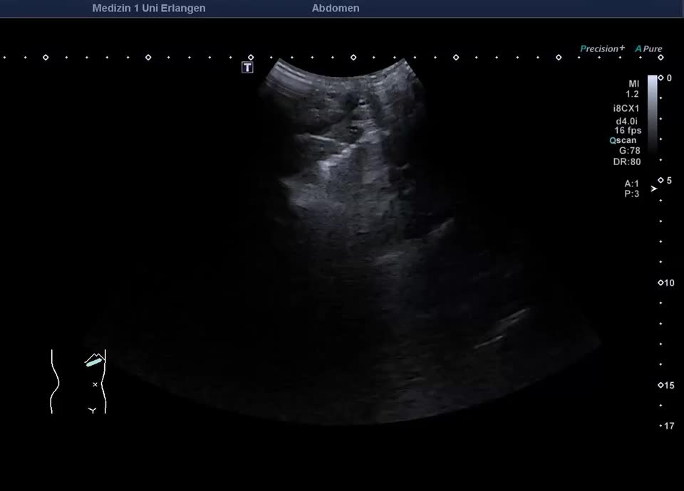
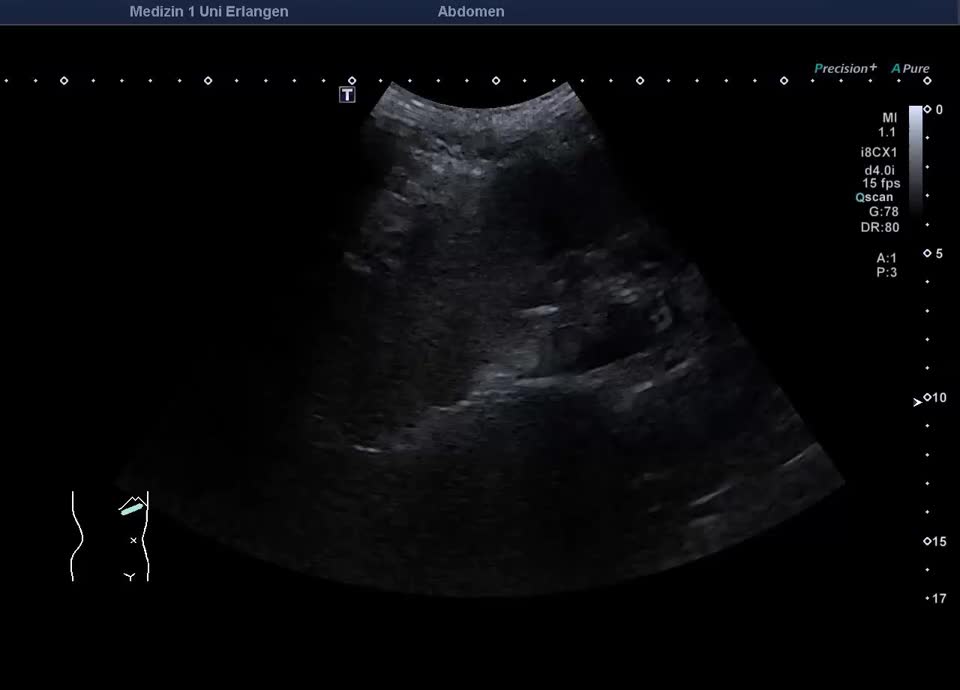
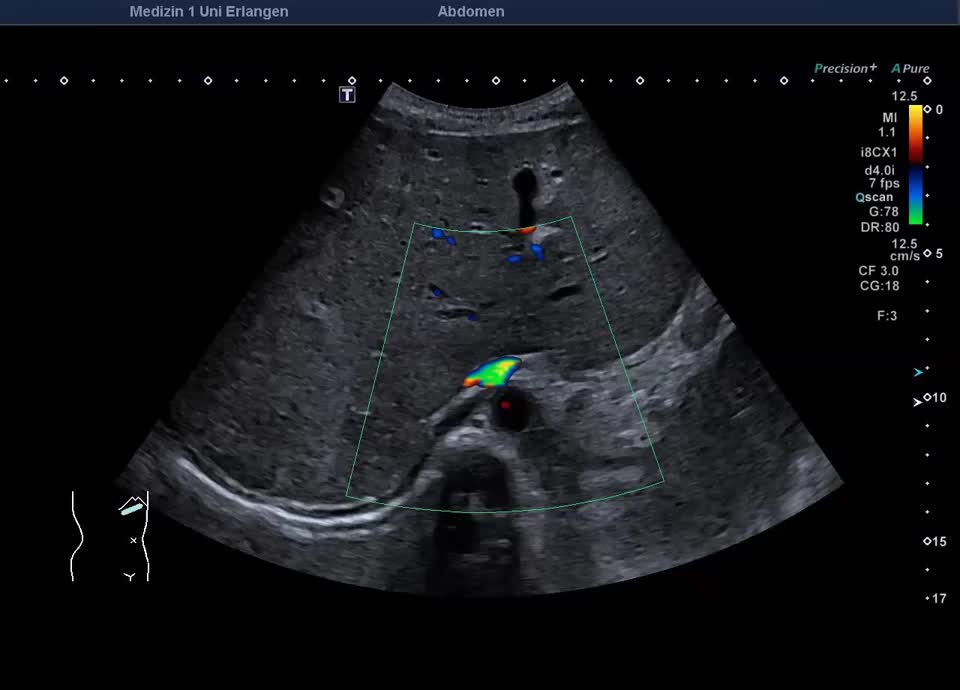
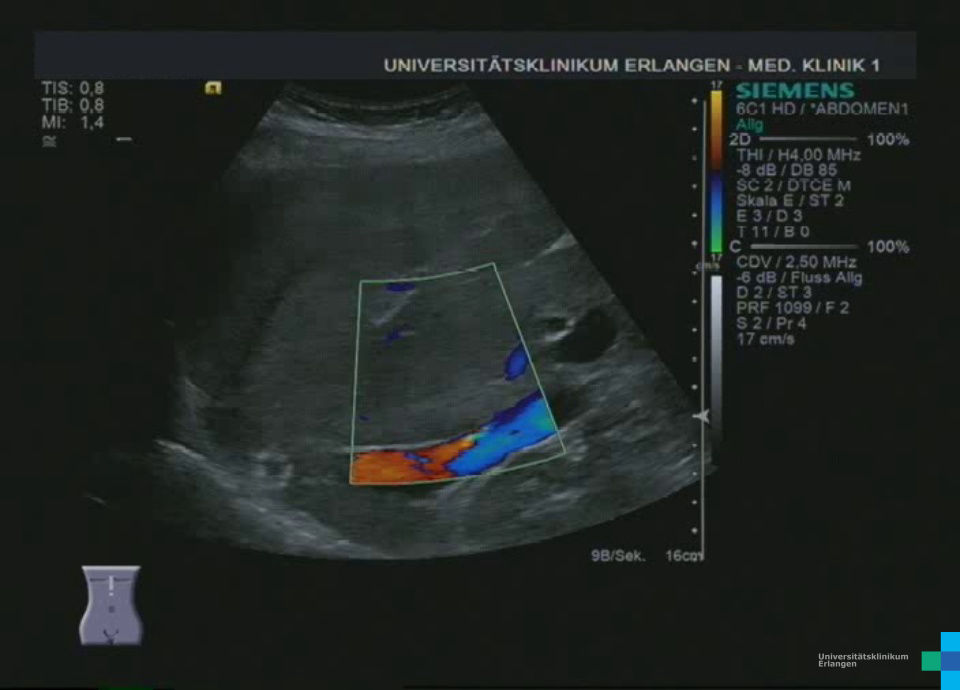
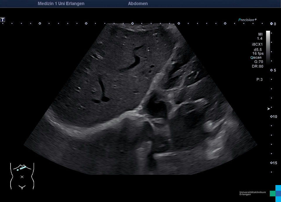
Case report: 37-year-old female patient, diagnosed with Budd-Chiari syndrome 03/25 externally, etiology unclear. Medical history: Crohn’s disease, HELLP syndrome (2022 during 2nd pregnancy). Sonographic findings upon admission show hepatomegaly with a rounded lower liver edge and hypertrophic caudate lobe and perihepatic ascites. The three hepatic veins are almost completely occluded (partially obliterated), intrahepatic venous collateralization through the hypertrophic caudate lobe to the inferior vena cava. The overall ultrasound imgae is consistent with chronic Budd-Chiari syndrome. Reduced portal vein flow before TIPSS placement with a maximum velocity of 13 cm/s. After successful TIPSS placement, the portal vein flow velocity increased to 40.6 cm/s.