

Liver abscess
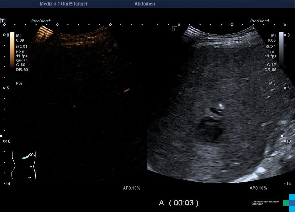
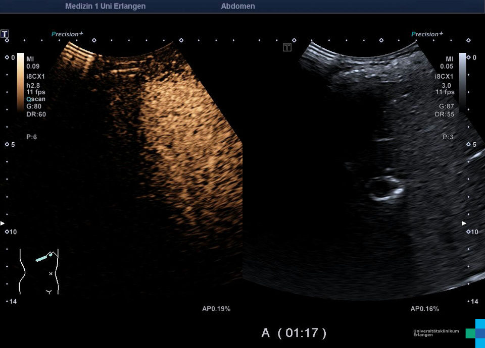
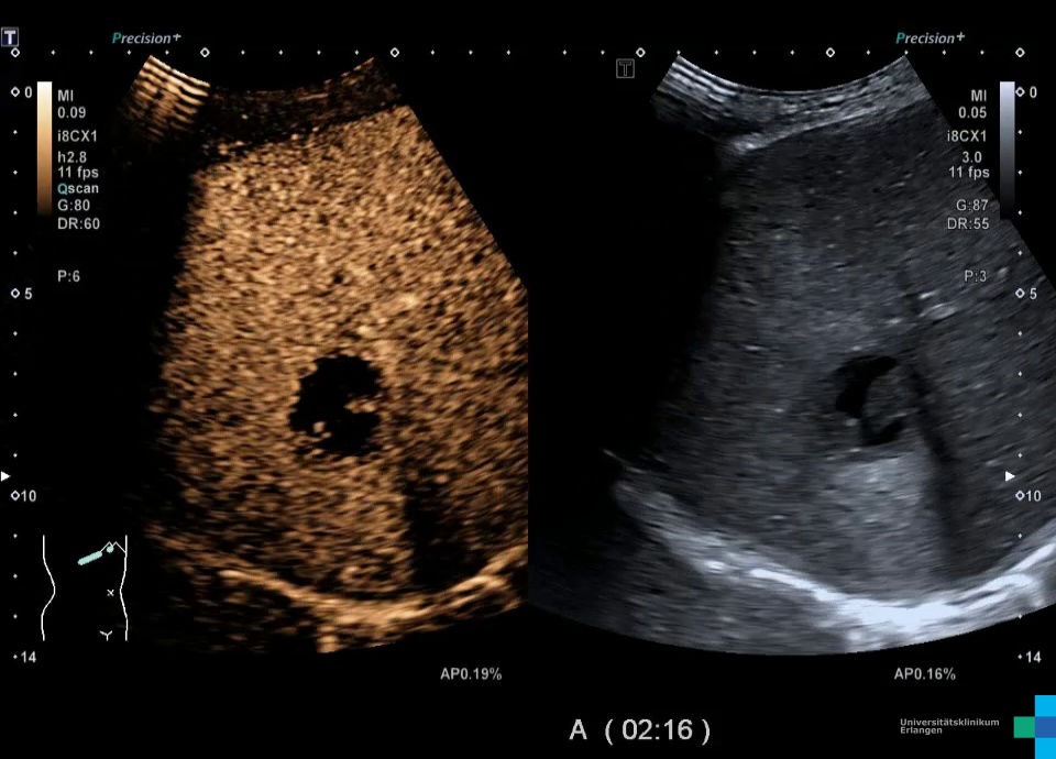
Case 1
Epicrisis: 54-year-old immunosuppressed patient following a kidney transplant. Currently presenting with sepsis due to a liver abscess caused by bile duct stenosis. 3.5 cm fluid-filled liver mass with echogenic internal echoes. On CEUS (arterial phase), the mass appears avascular (non-enhanced) with a very narrow hyper-enhanced rim (reactive hyperemia, visible only in the arterial phase). In the portal venous phase or late phase, the lesion remains completely avascular (non-enhanced). Vancomycin-resistant enterococci were found in the abscess aspirate.


Case 2
Epicrisis: 35-year-old patient after respiratory infection with pleural effusion and pneumonia. CT-graphically, a liver lesion was found, with a suspicion of liver abscess. In CEUS the liquid parts are non-enhancing, the abscess capsule shows arterial hyperenhancement.


Case 3
Epicrisis: 69-year-old patient with past resection of a cholangiocellular carcinoma and postoperative abscess. Ultrasound shows an encapsulated liquid mass in segment VII. CEUS shows arterial hyperemia in the surrounding liver tissue. In the portal venous and late phase demarcation of the liquid parts without enhancement.


