
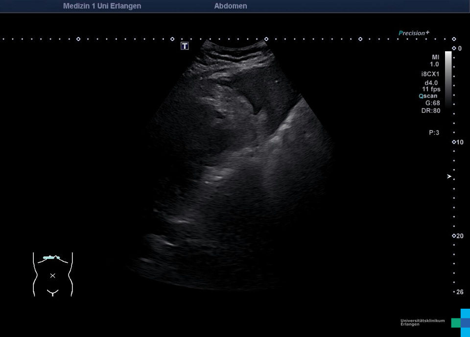
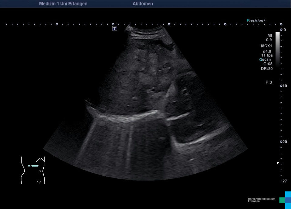
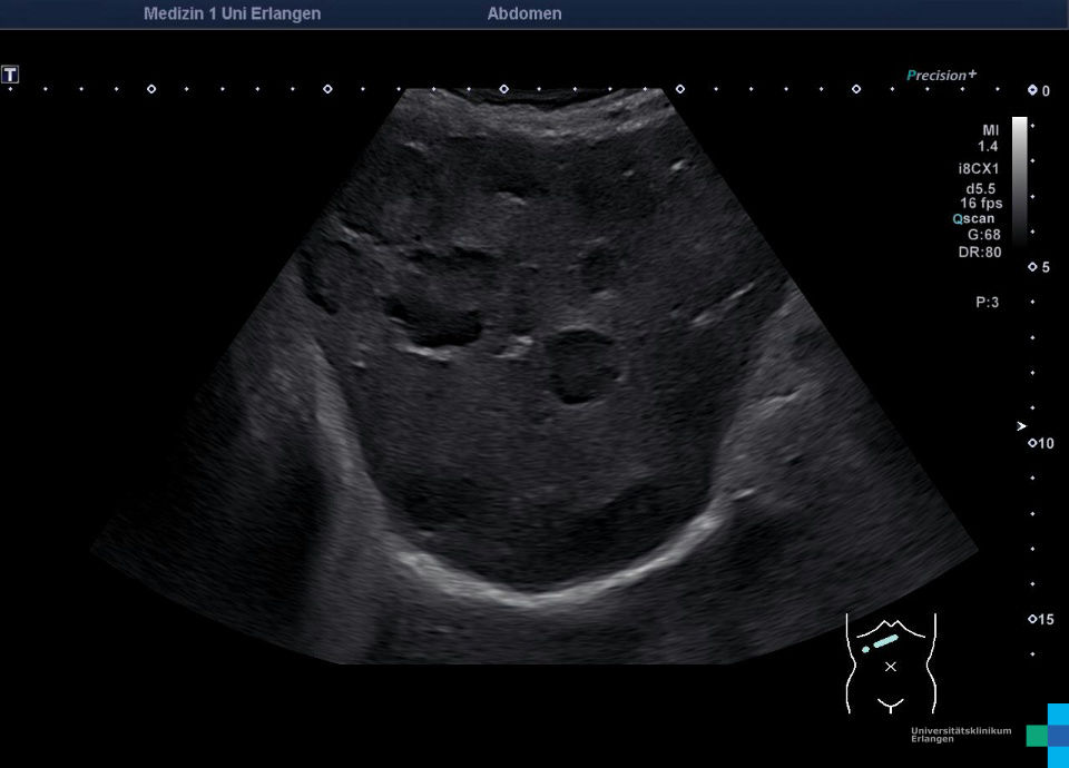
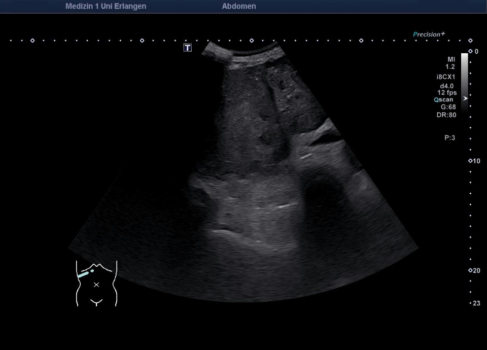
Liver metastases


Kasuistik
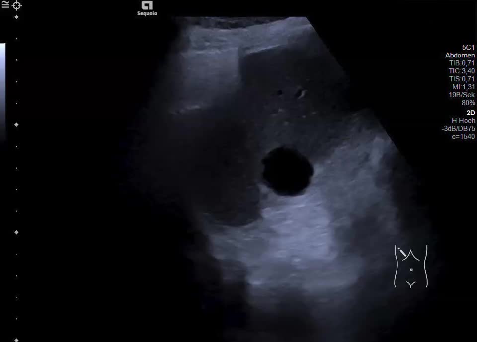
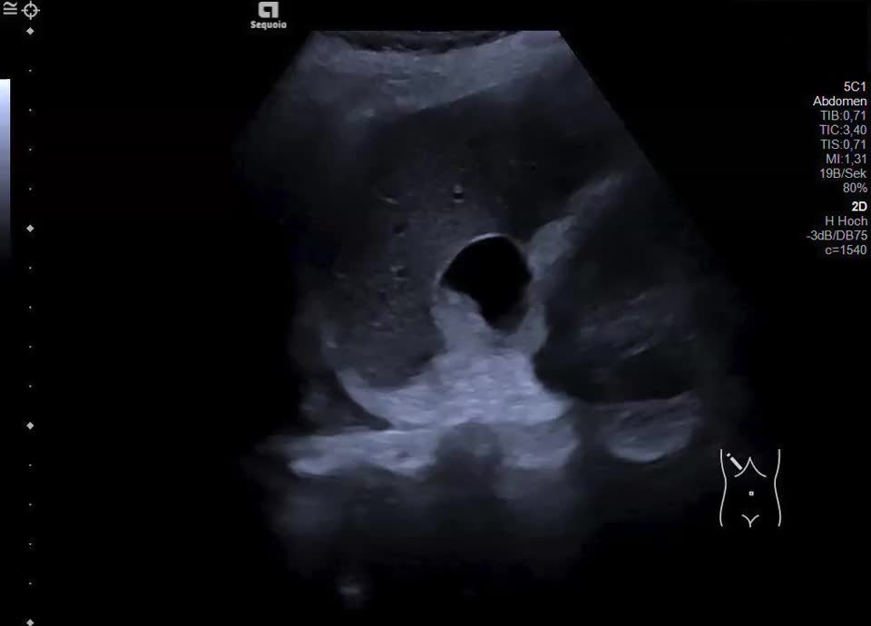
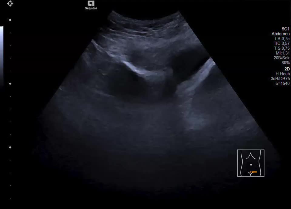
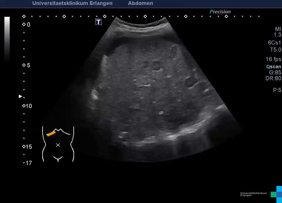
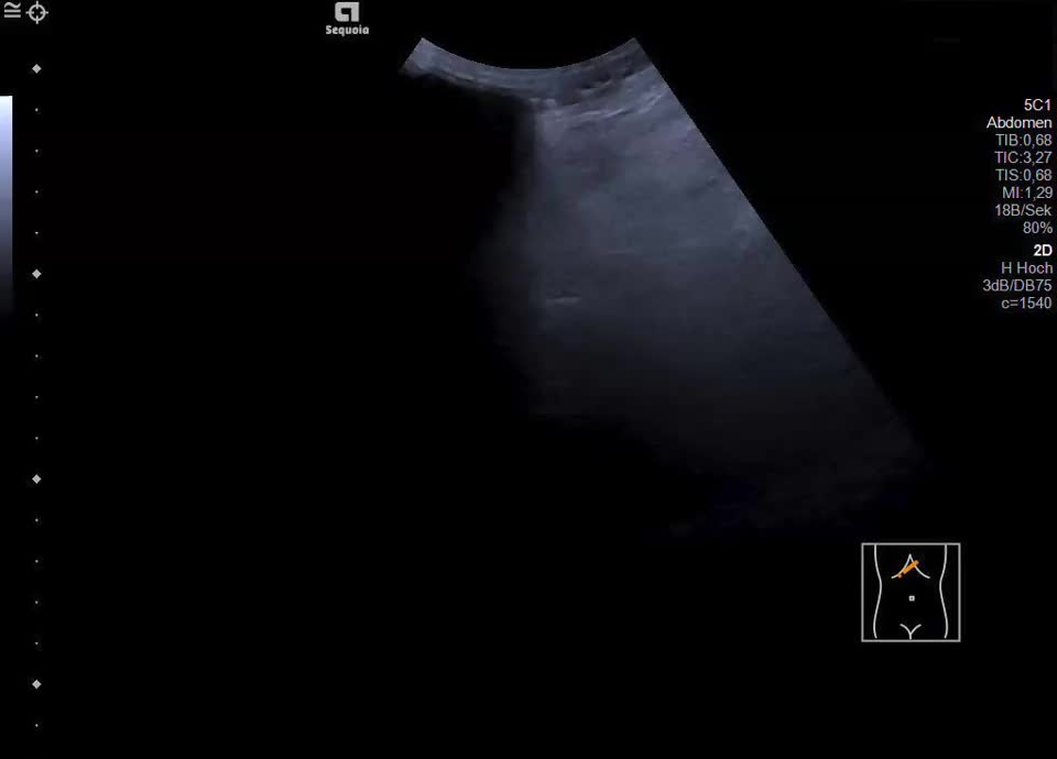
A 61-year-old female patient with suspected antiphospholipid syndrome, status post multiple cerebral infarctions and pulmonary embolism of unclear etiology, underwent abdominal ultrasound evaluation. Ultrasound demonstrated a subcapsular cystic lesion in hepatic segment VI with a continuous capsular echo and an intralesional hyperechoic solid component. The findings are not consistent with a simple cyst but are suspicious for a cystic-solid metastasis. On CEUS, the solid component showed contrast enhancement (s.a. CEUS folder). Additionally, a nodular peritoneal thickening in the upper abdomen and a cystic ovarian tumor with solid components in the lower abdomen are present. In summary, the findings are highly suggestive of a hepatic and peritoneal metastasizing ovarian carcinoma.