

Cholestasis
-
 Dilated bile ducts with stents (video)
 -
 Dilated bile ducts with sludge (video)
 -
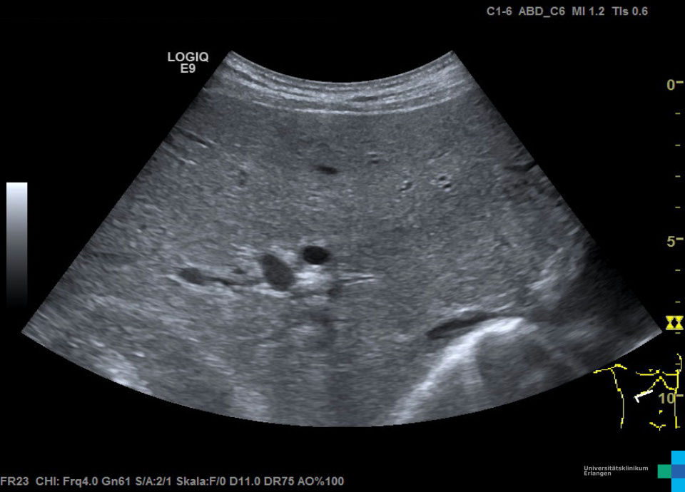
 Intrahepatic bile duct dilation
 -
 Intrahepatic bile duct dilation
 -
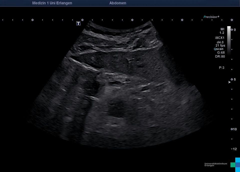
 Dilated bile duct due to choledocholithiasis
 -
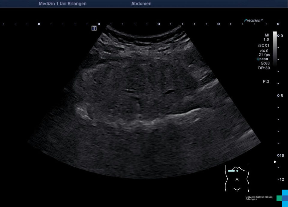
 Ectatic common hepatic duct with cystic duct stump after cholecystectomy
 -
 Cholestasis due to pseudocyst
 -
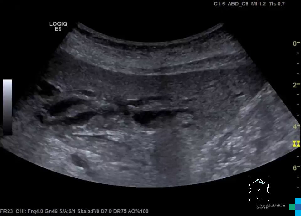
 Dilated bile ducts
 -
 Dilated bile ducts (video)
 -
 PTCD (2D-mode video)
 -
 PTCD after contrast agent injection (Video)
 -
 Contrast agent in small bowel (video)
 -
 Choledocholithiasis
 -
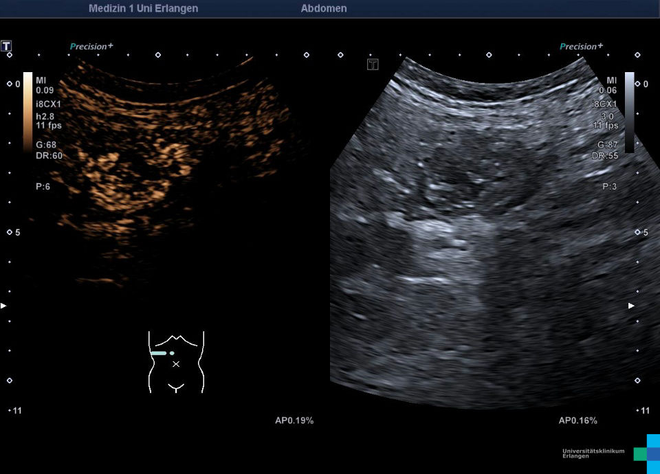
 Intrahepatic cholestasis
 -
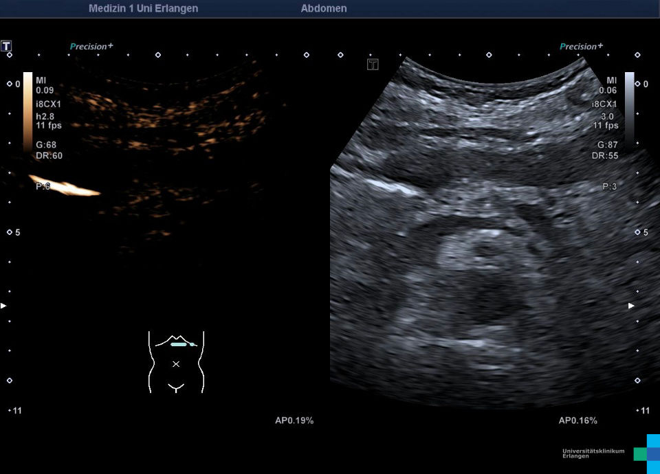
 Intrahepatic cholestasis (color doppler image)
 -
 Intrahepatic cholestasis (video)
 -
 Cholestasis (video)
 -
 Cholestasis
c
Hepatikusgabel mit erweitertem Ductus hepaticus dexter und sinister (jeweils 6 mm) und erweitertem wandverdickten DHC (18 mm) mit einliegenden DHC-Stents und Sludge
Erweiterte Gallenwege mit Sludge
Dilatation der intrahepatischen Gallenwege (Doppelflinten)
Dilatation der intrahepatischen Gallenwege (Doppelflinten)
Erweiterter DHC (B) bei Choledocholithiasis, in der Papille vateri fixiertes Konkrement (A)
Ektatischer DHC mit abgesetztem Zystikusstumpf bei Z.n. Cholezystektomie
Ultraschall, Ultraschallatlas, Ultraschallbilder, Ultraschallvideos, Sonographie, Sonographieatlas, Sonographiebilder, Sonographievideos, Kontrastmittelultraschall, Kontrastmittelsonographie, Medizinische Klinik 1, Uniklinik, Universitätsklinikum