

HCC without liver cirrhosis (series of images/videos)


Epicrisis:
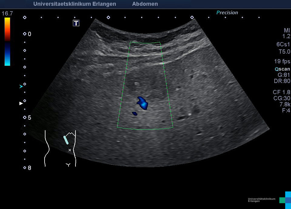
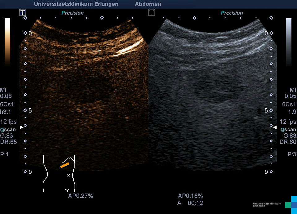
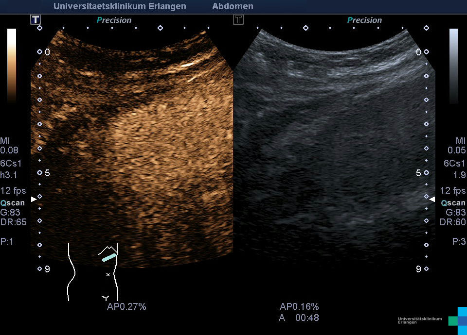
65-year-old patient with fatty liver (diabetes mellitus, arterial hypertension) with hypoechoic mass in liver, incidental finding by primary care physician. An out of house performed CT and MRI can’t supply additional information. CEUS shows a vascularization pattern suspicious for an HCC (arterial hyperenhancement with beginning contrast agent wash-out after 3 minutes). Histological examination confirms an HCC (G2), tissue doesn’t show signs of liver cirrhosis and no sign of liver fibrosis.
For better visualization due to didactic reasons the playback speed got reduced to 75%.