

Crohn’s Disease
-
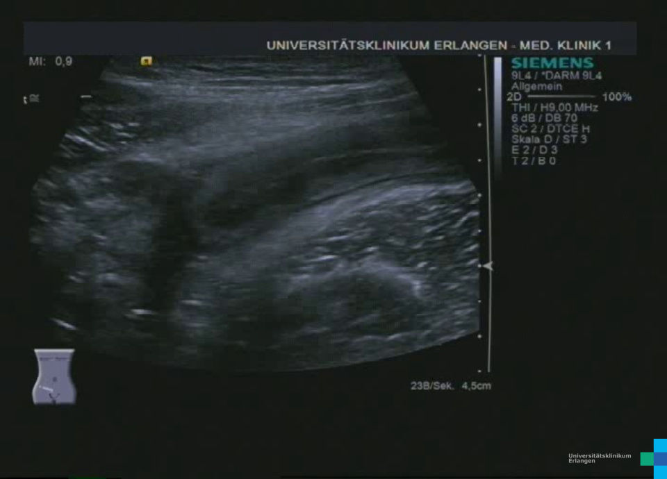
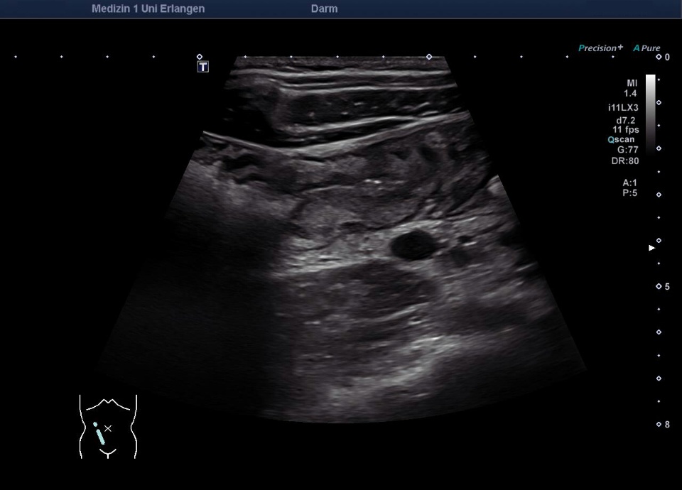
 Thickened wall of the terminal ileum
 -
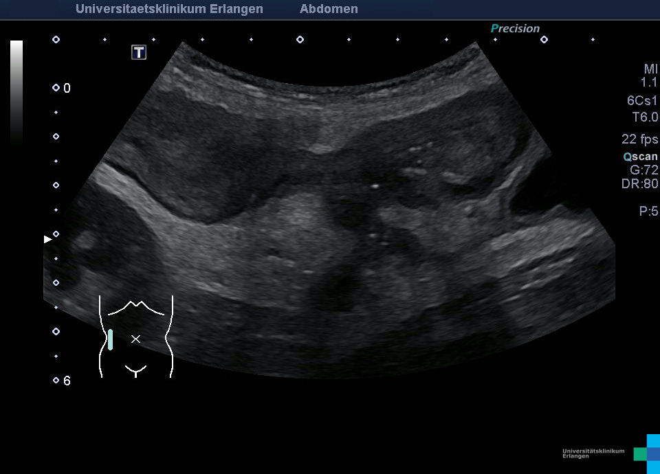
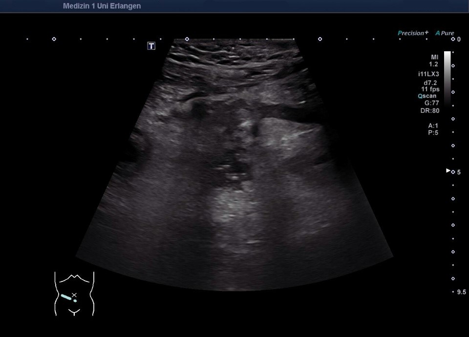
 Elongated acute terminal ileitis (video)
 -
 Elongated acute terminal ileitis (video)
 -
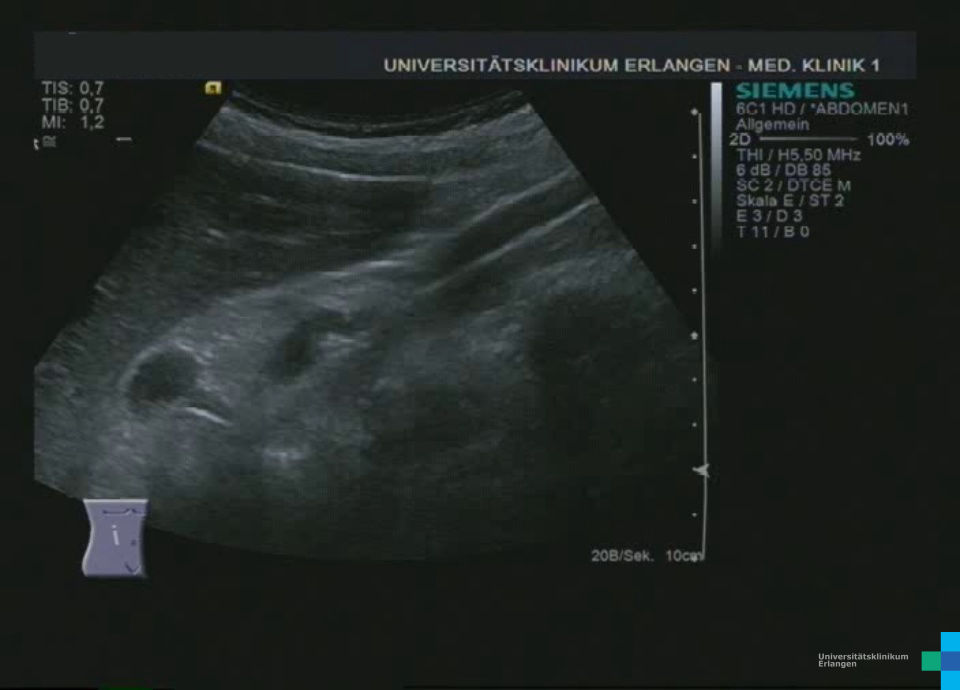
 Fistula in patient with Crohn's disease (video)
 -
 Fistula in patient with crohn's disease
 -
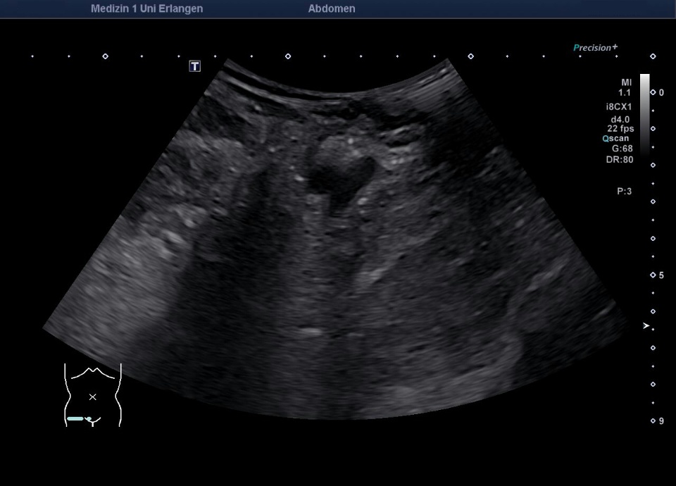
 Inflammation of the terminal ileum
 -
 Ureteral obstruction caused by crohn's disease with abscesses
 -
 Ureteral obstruction caused by crohn's disease (video)
 -
 Ureteral obstruction caused by crohn's disease with abscesses
 -
 Ileitis (M. Crohn)
 -
 Ileitis (M. Crohn, color doppler)
 -
 Ileitis (M. Crohn, SMI)
 -
 Ileitis (M. Crohn, video)
 -
 abscess in Crohn's ileitis
 -
 abscess in Crohn's ileitis
 -
 abscess in Crohn's ileitis (Video)
 -
 ileitis terminalis in crohn's disease
 -
 Ileitis terminalis in crohn's disease (video)
 -
 Fistulating Crohn's disease (video)
 -
 Fistulating Crohn's disease
Terminales Ileum wandverdickt bei Morbus Crohn: * = Muskularis, ** = Submukosa, *** = Mukosa (hochfrequenter Linearschallkopf)
Langstreckige akute Ileitis terminals bei Morbus Crohn (Bezeichnung der Strukturen folgt im nächsten Bild; hochfrequenter Linearschallkopf)
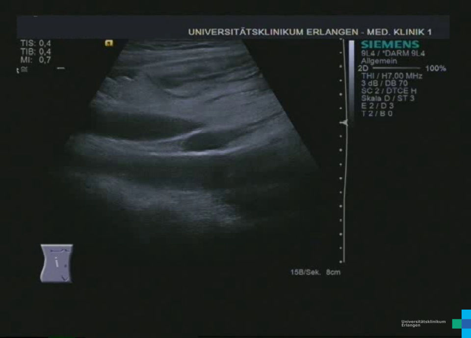
Langstreckige akute Ileitis terminals bei Morbus Crohn (hochfrequenter Linearschallkopf)
Fisteln als Komplikation eines Morbus Crohn mit echoreicher Umgebungsreaktion (Bezeichnung der Strukturen folgt im nächsten Bild)
Fisteln als Komplikation eines Morbus Crohn mit echoreicher Umgebungsreaktion (* = Fistel)
Wandverdicktes terminales Ileum – murale Hyperämie im Farb-Doppler (hochfrequenter Linearschallkopf)
Ultraschall, Ultraschallatlas, Ultraschallbilder, Ultraschallvideos, Sonographie, Sonographieatlas, Sonographiebilder, Sonographievideos, Kontrastmittelultraschall, Kontrastmittelsonographie, Medizinische Klinik 1, Uniklinik, Universitätsklinikum, Erlangen