

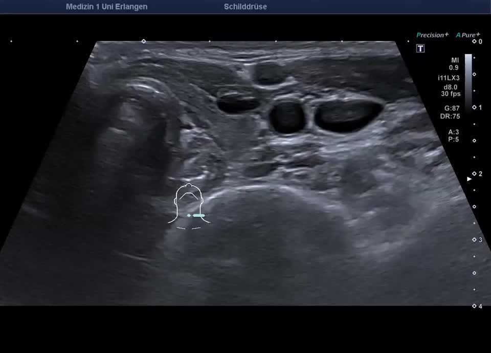
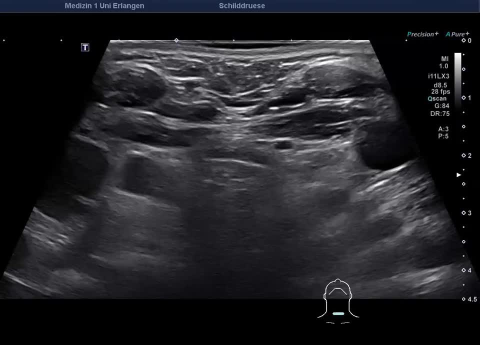
Autoimmune thyroiditis


Case report:
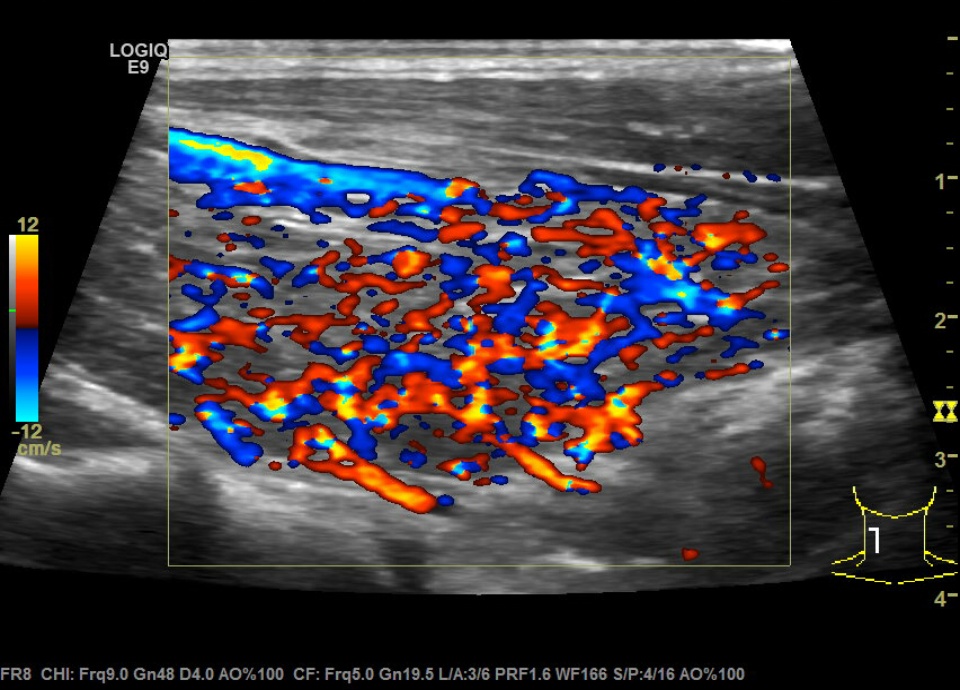
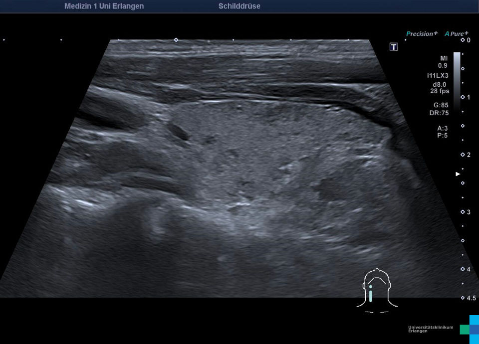
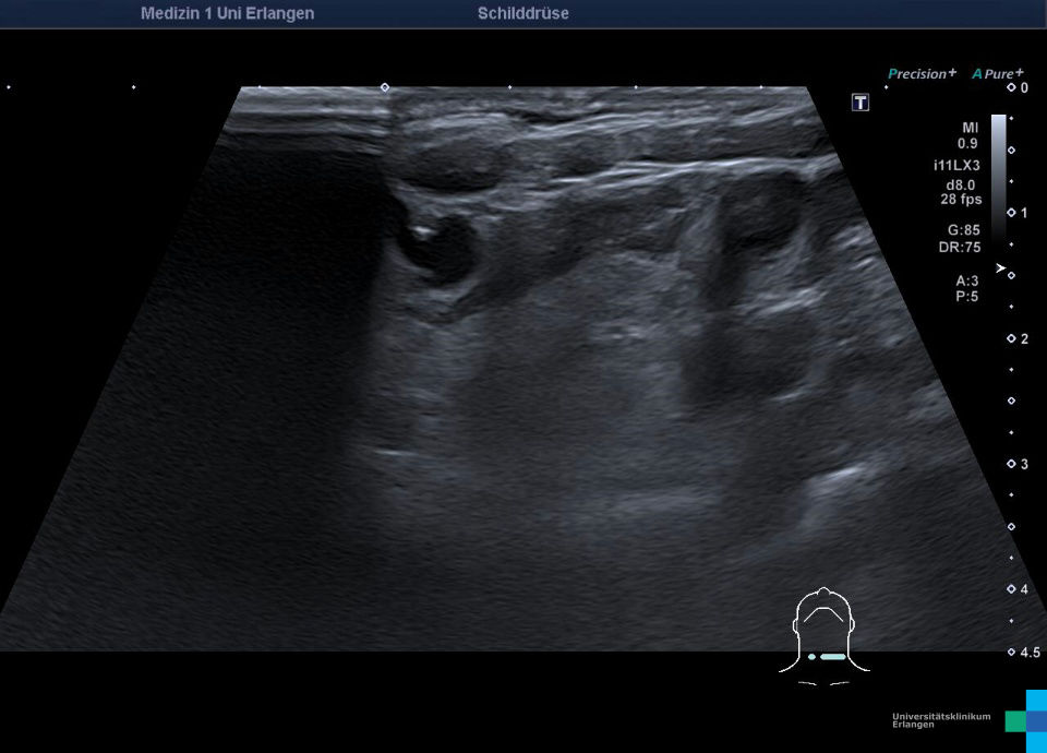
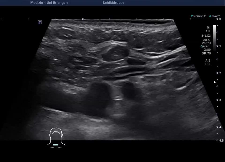
36-year-old patient with tremor, tachycardia, exophthalmos, and weight loss. Thyroid ultrasound shows bilaterally hypoechoic, inhomogeneous parenchyma with a total volume of 20.5 ml. Color Doppler sonography shows hypervascularization. Laboratory results: basal TSH decreased, T3/fT4 elevated, TRAK positive, consistent with Graves’ disease. Symptoms resolved under treatment with thiamazole and propranolol. At follow-up two years later, sonography showed a hyperechoic minimally inhomogeneous parenchyma with normal volume (15 ml).


