
Von Meyenburg complexes
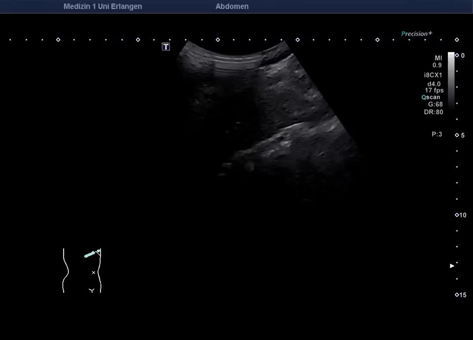
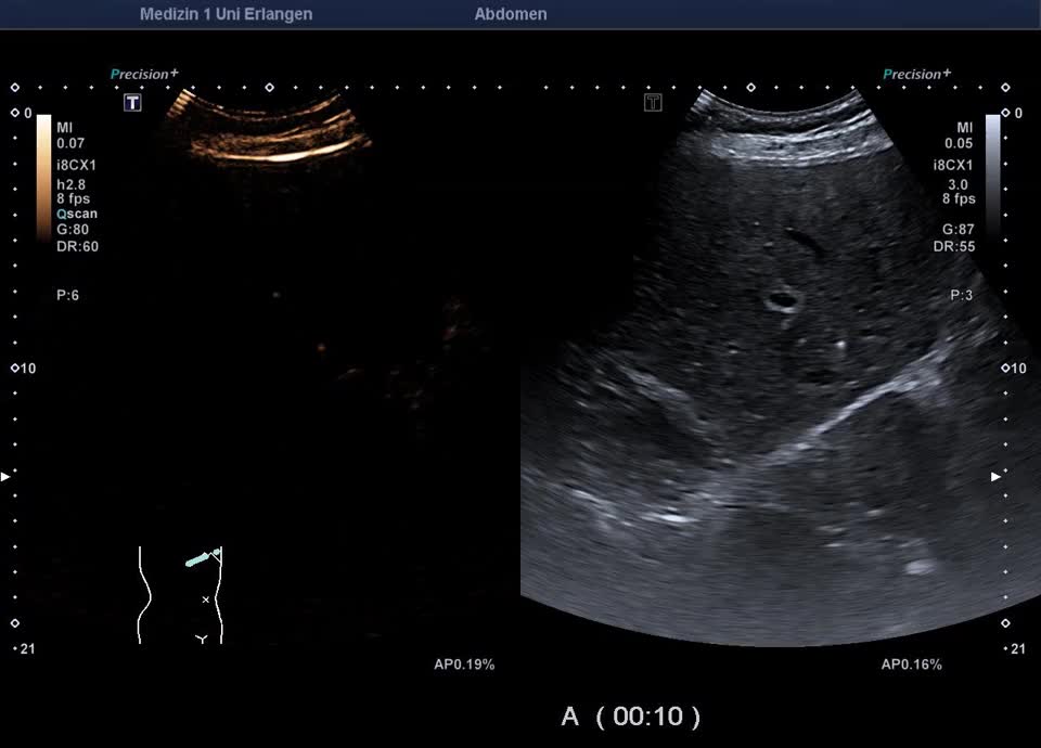
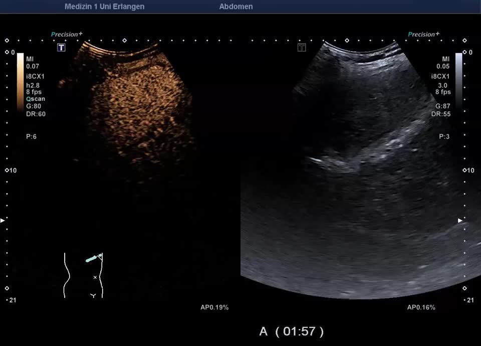
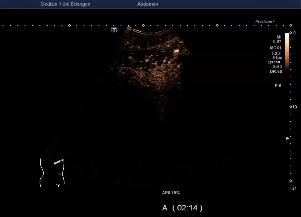
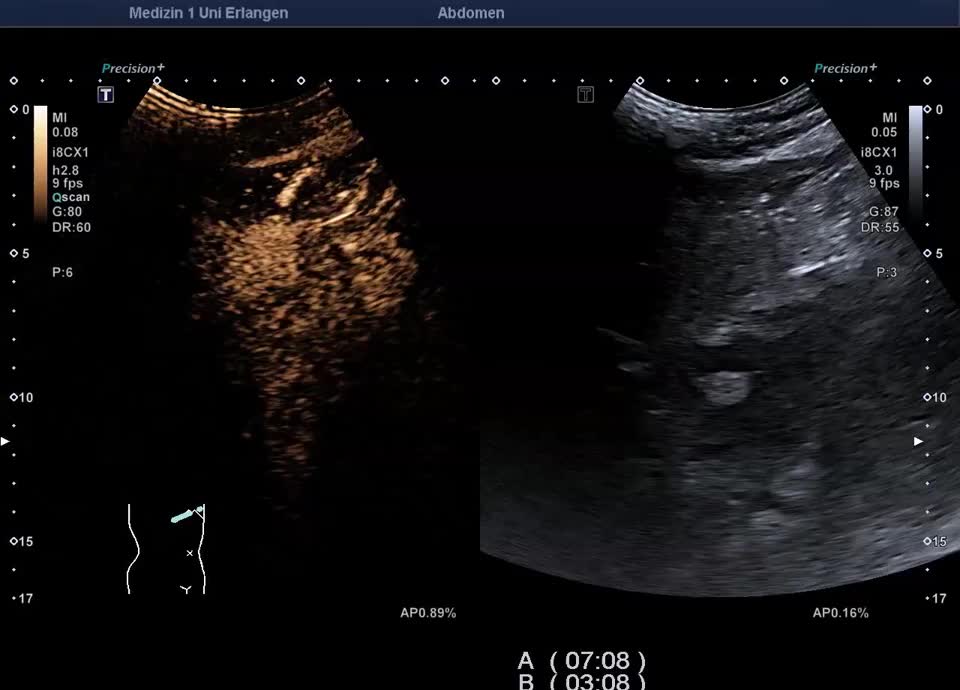
Case report: 66-year-old male patient with a long-standing elevation of γ-GT since adolescence was referred for sonographic evaluation to exclude cholangiocellular carcinoma. B-mode ultrasound revealed an inhomogeneous liver parenchyma with diffusely distributed small comet-tail artifacts in the right liver lobe. The sonographic appearance was consistent with multiple biliary hamartomas (Von Meyenburg complexes). These consist of cysts, lined with biliary epithelium, and connective tissue, occur singly or in multiples, and typically measure 0.5–15 mm. Sonographically, they appear as echogenic nodules. To exclude malignancy-suspicious focal lesions, a contrast-enhanced ultrasound (CEUS) was performed. On CEUS, the liver parenchyma showed homogeneous enhancement in the portal venous and late phases. The small hyperechoic nodules are isocontrasted (excluding malignancy). The small cysts are unenhanced in any perfusion phase. In correlation with the imaging findings and the known γ-GT elevation, there was no evidence of malignant liver disease. A single follow-up sonographic examination was scheduled.