
Thyroid nodule




Case report:
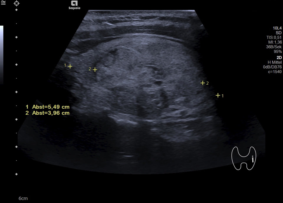
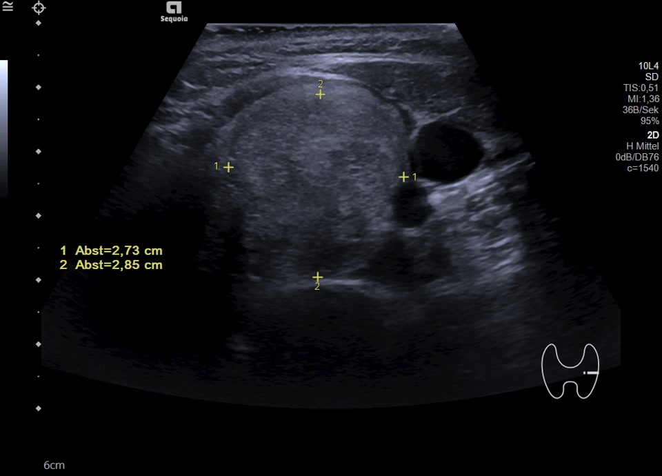
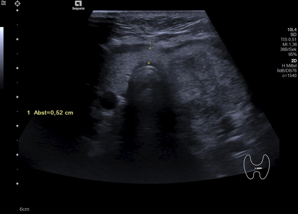
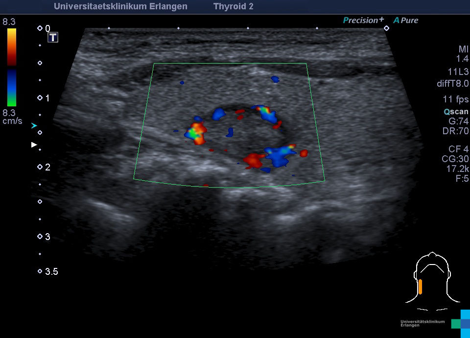
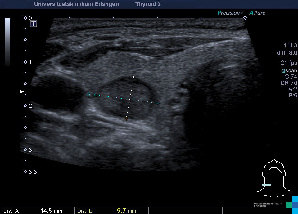
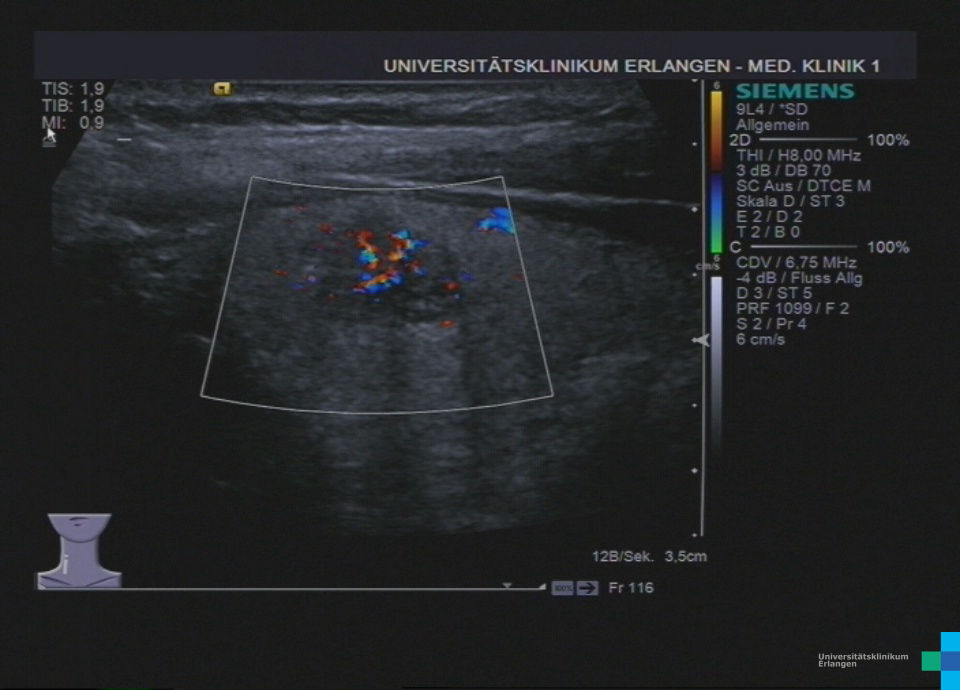
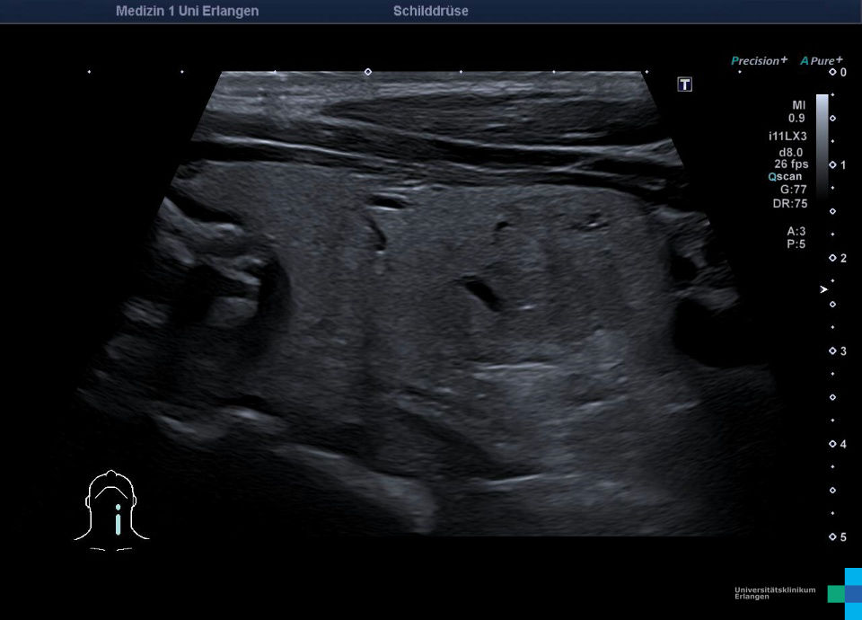
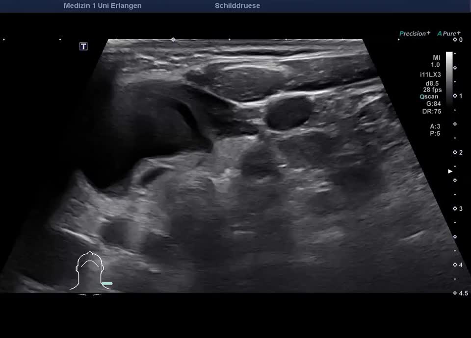
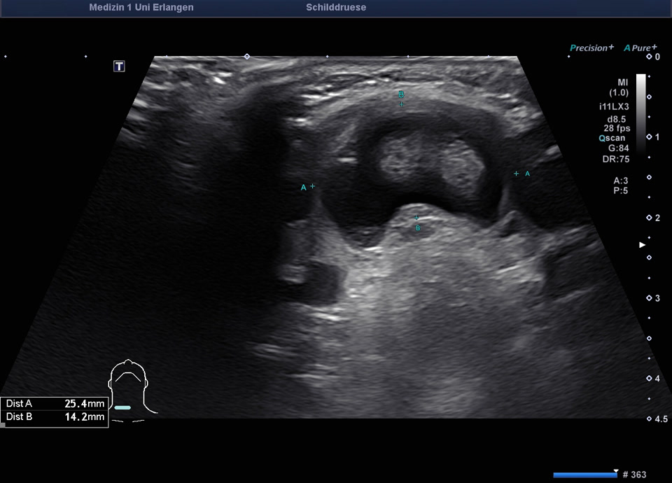
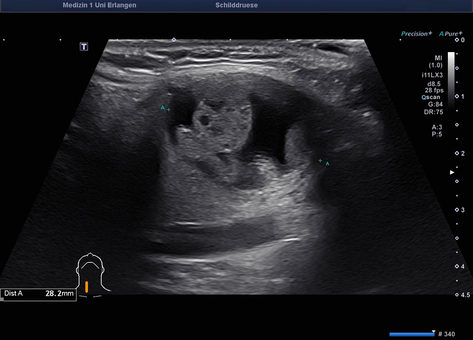
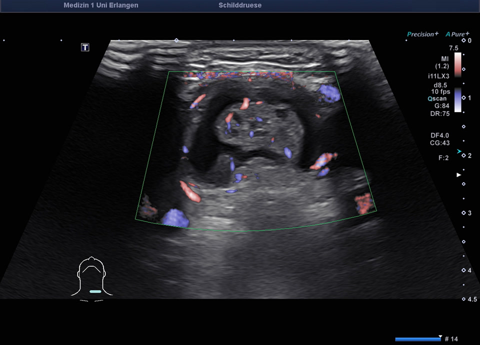
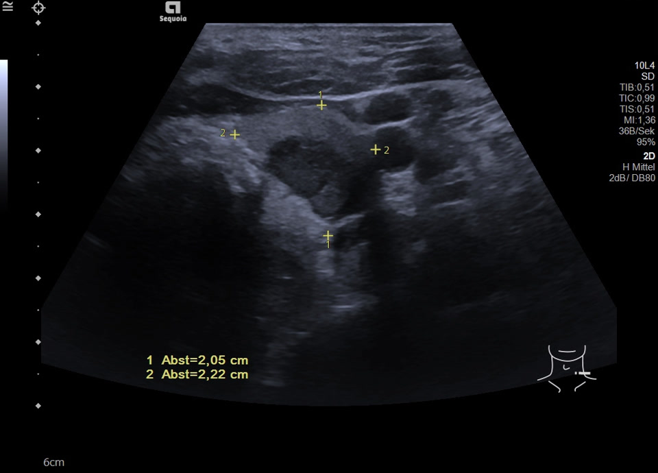
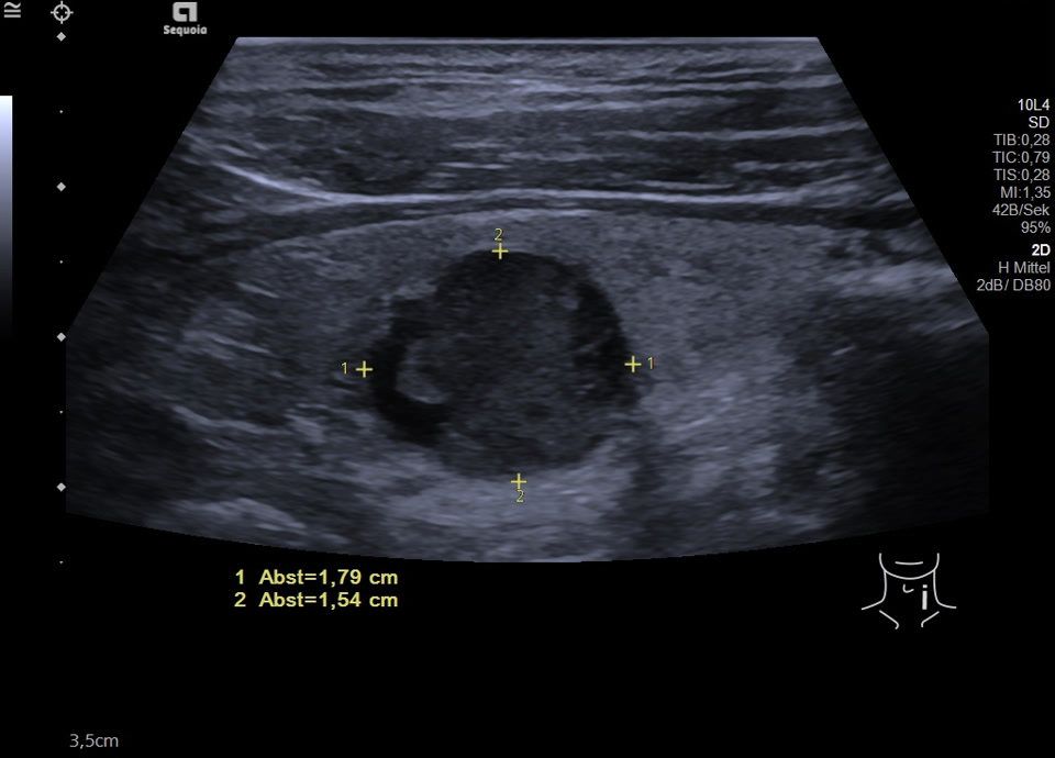
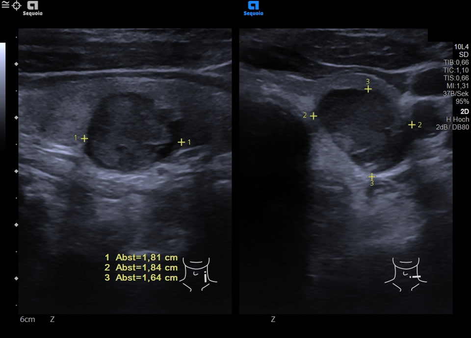
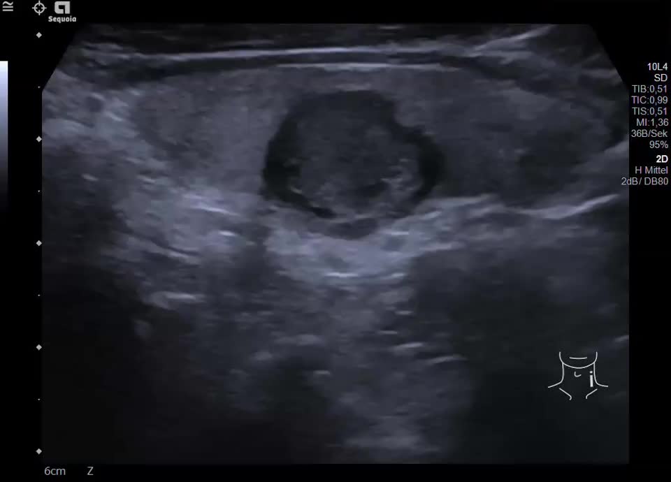
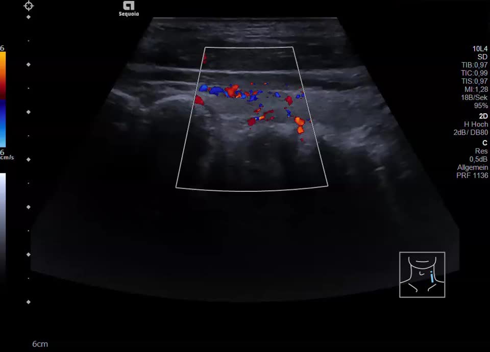
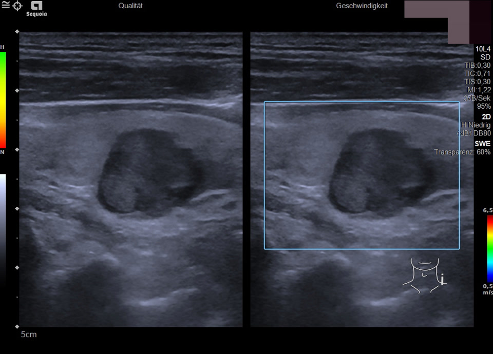
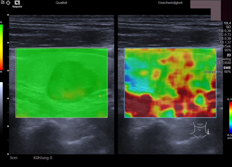
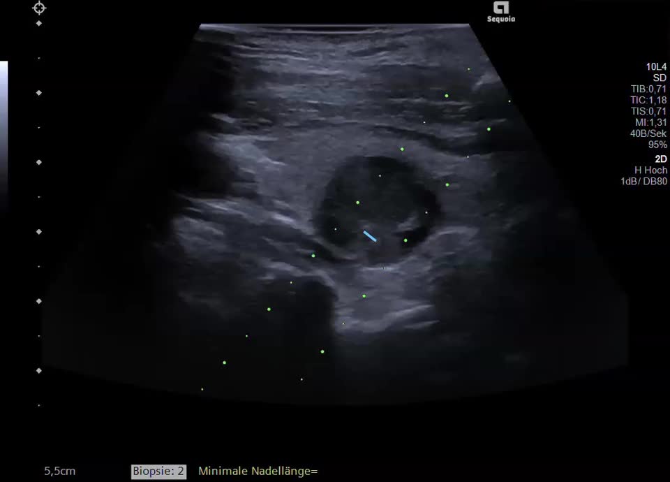
A 78-year-old patient with an incidental finding of a hypodense thyroid lesion on cardiac CT performed prior to planned aortic valve surgery. Ultrasound examination revealed a thyroid gland of normal size with a 2-cm hypoechoic nodule (EU-TIRADS 4); color Doppler imaging showed no significant intranodular blood flow. Elastometry showed increased tissue stiffness. The TSH level was within the normal range, and there were no clinical indications of thyroid disease. An ultrasound-guided core needle biopsy and a fine-needle aspiration with cytological analysis (cytospin) were performed. Histologically, a follicular nodule was identified. Pathologist’s comment: Based on the biopsy, a follicular neoplasm cannot be reliably distinguished from an autonomous adenoma. Thyroid scintigraphy should be performed for functional evaluation (to detect an autonomous adenoma). Due to the prior administration of contrast medium (cardiac CT), a thyroid scintigraphy is only possible after ≥8 weeks. At present, treatment of the severe aortic stenosis is prioritized. If scintigraphy shows no evidence of autonomy, surgical evaluation via diagnostic hemithyroidectomy should be considered due to the risk of a follicular neoplasm. Alternatively, ultrasound follow-up at 6 and 12 months may be performed.
Case report:
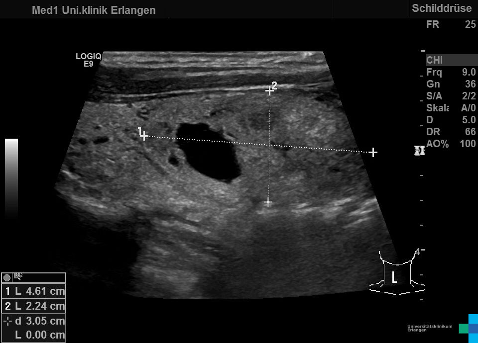
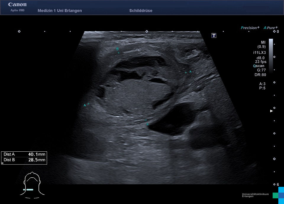
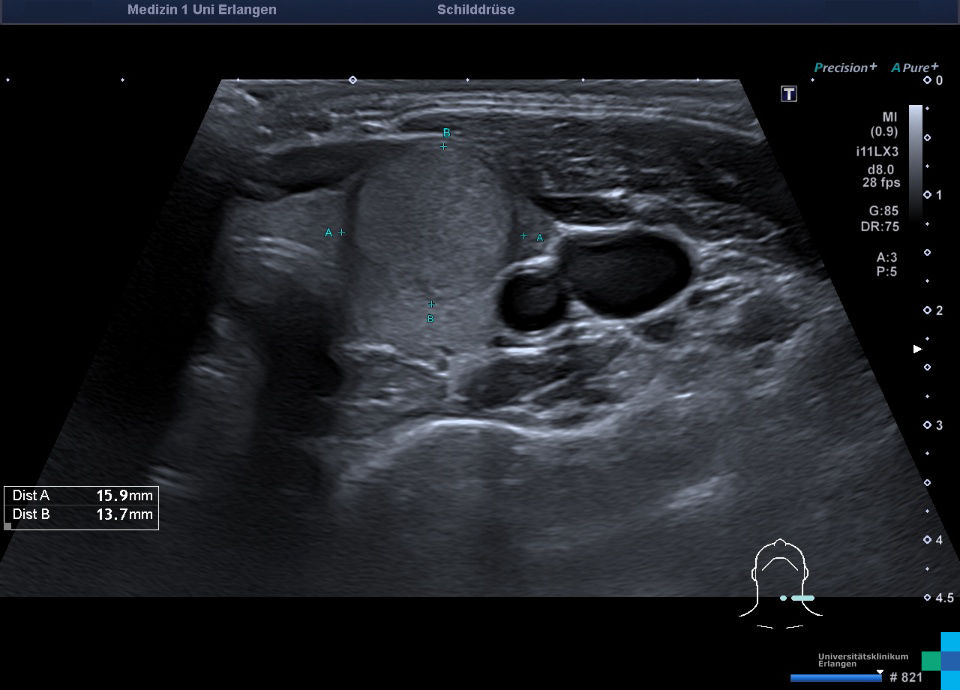
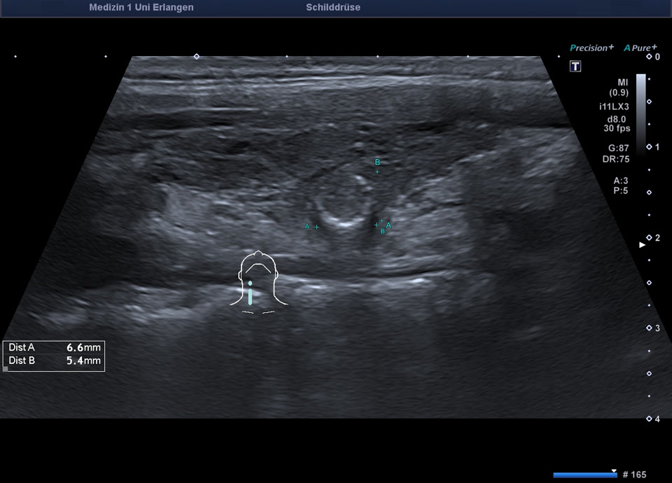
A 43-year-old female patient presented with a sensation of pressure on the left side of the neck as well as dysphagia. Laboratory evaluation revealed normal TSH, fT3, and fT4 levels. Ultrasound demonstrated enlargement of the left thyroid lobe caused by an inhomogeneous solid nodule that was smoothly marginated and isoechoic relative to the surrounding thyroid parenchyma. No sonographic features of malignancy were identified (->EU-TIRADS 3). The remaining thyroid parenchyma appeared unremarkable. Due to the clinical symptoms, thyroid scintigraphy for functional characterization as well as surgical resection of the symptomatic nodule were recommended.