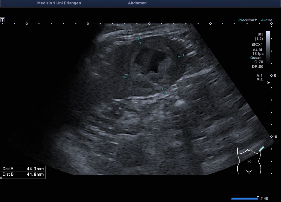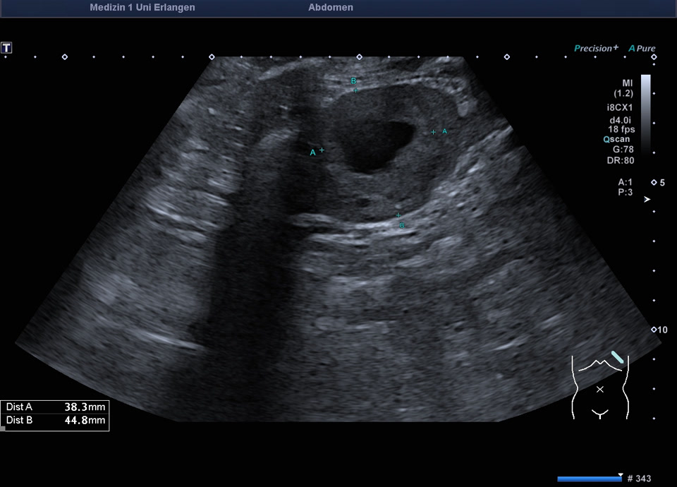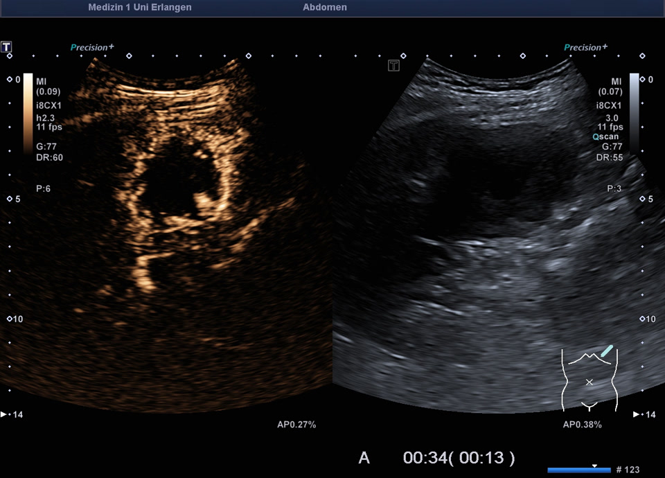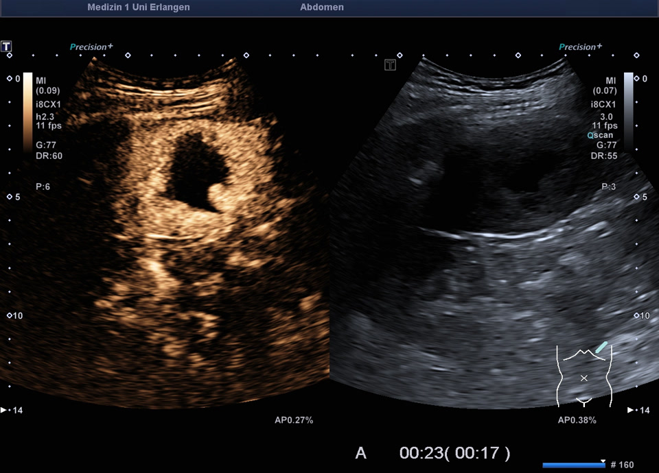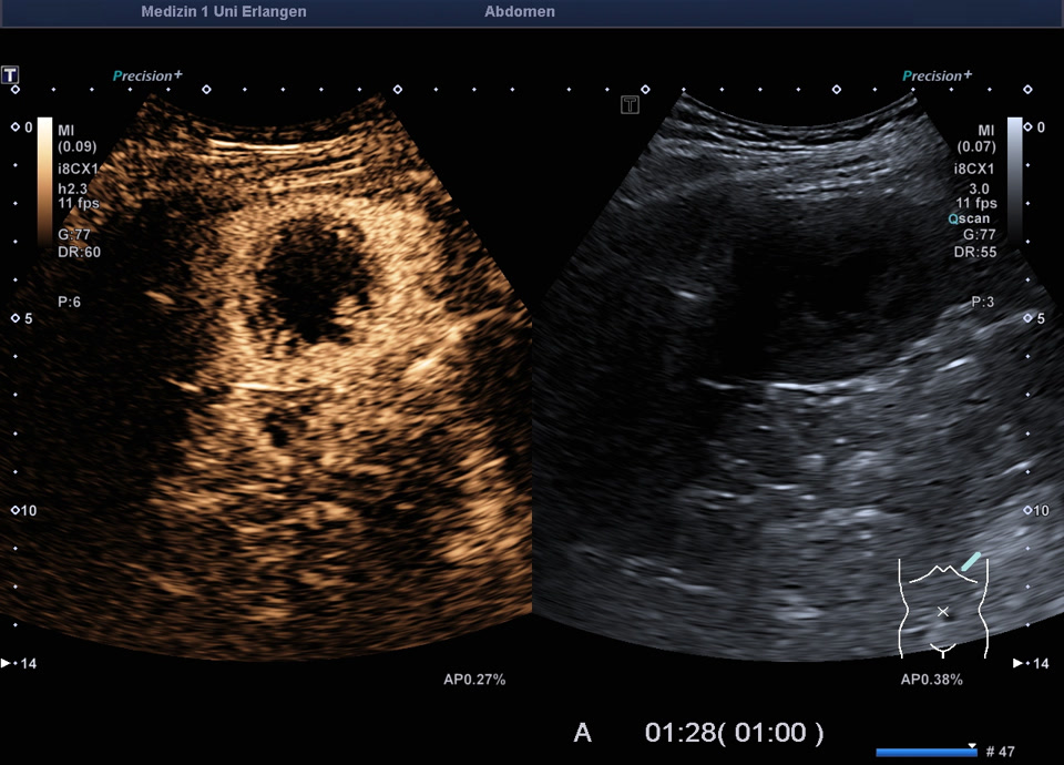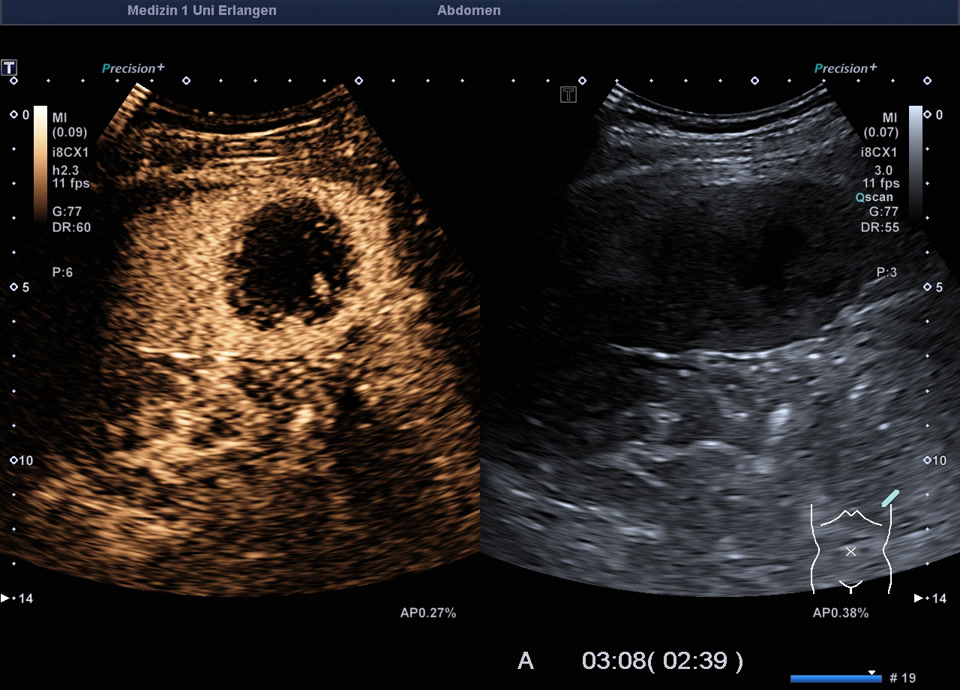

Splenic metastasis (melanoma)
Case report: A 75-year-old patient with malignant melanoma. A CT scan revealed enlarged mesenteric lymph nodes and a splenic lesion. Upon sonography, the lesion appears as a heterogeneous hyperechoic mass with central areas of fluid. Contrast-enhanced sonography shows arterial hyperenhancement of the lesion with contrast washout in the late phase. The central components do not take up contrast medium. Based on the overall findings, a splenic metastasis of the melanoma with central necrotic components is suspected.