Porphyria cutanea tarda 2 (Liver)
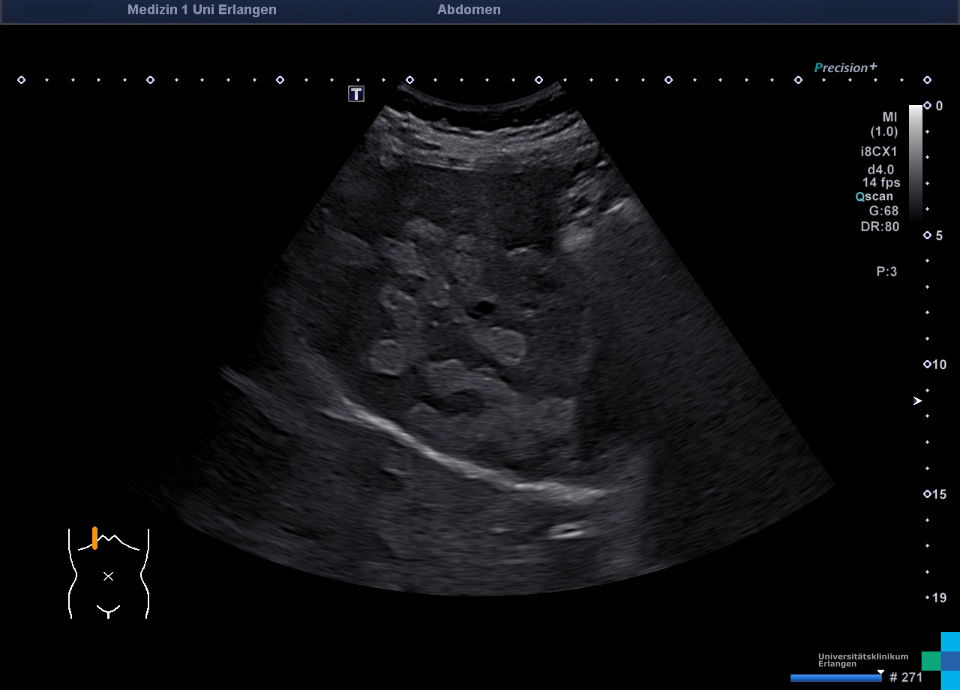
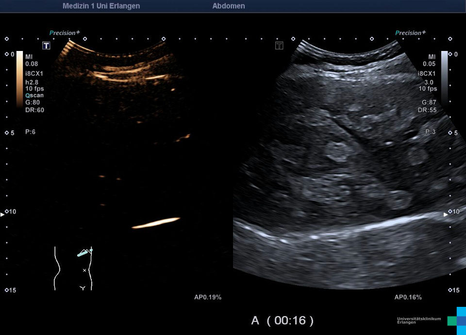
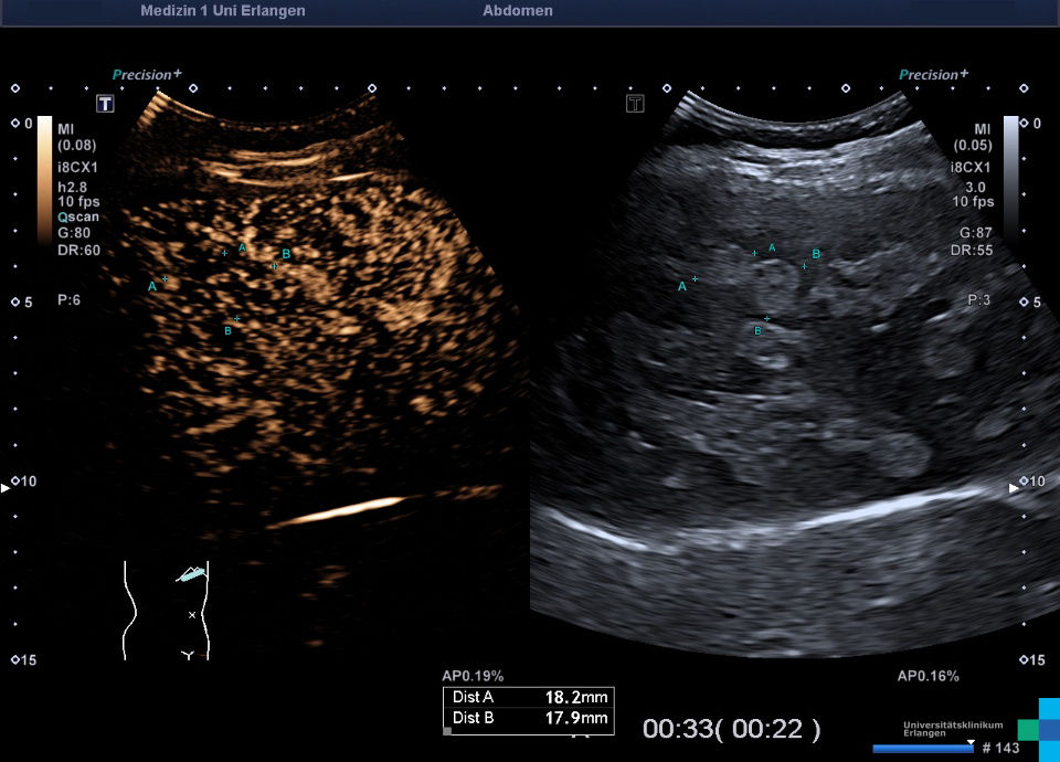
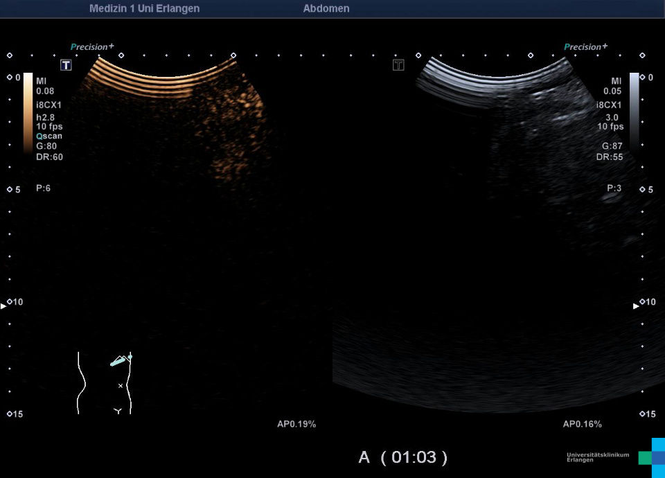
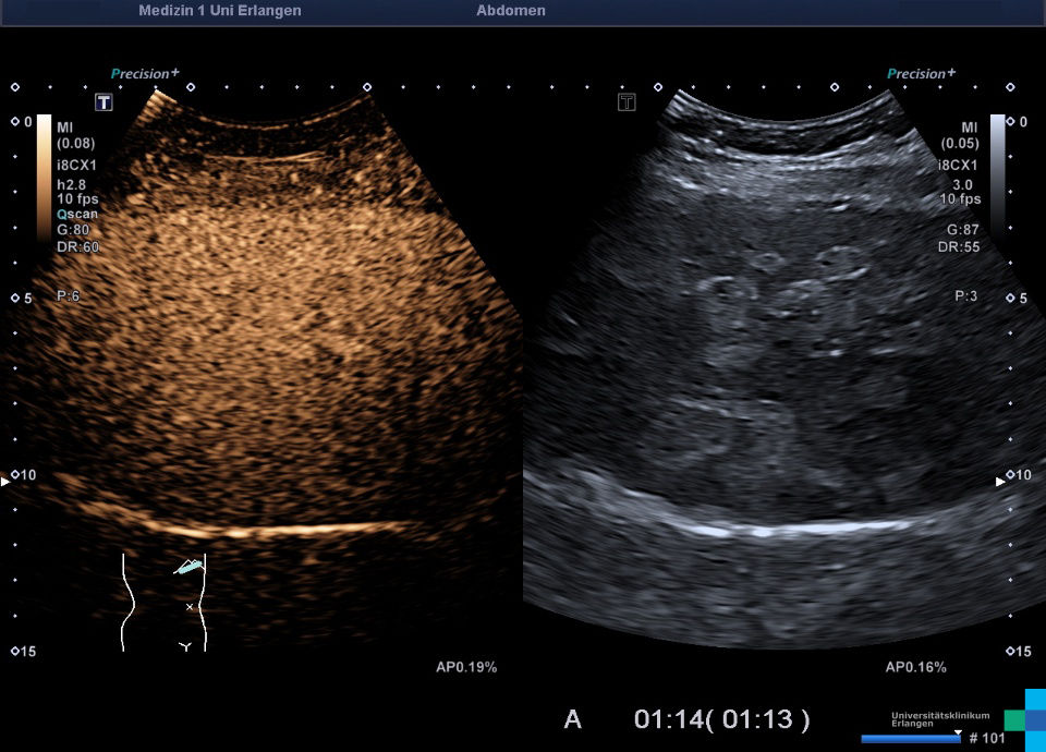
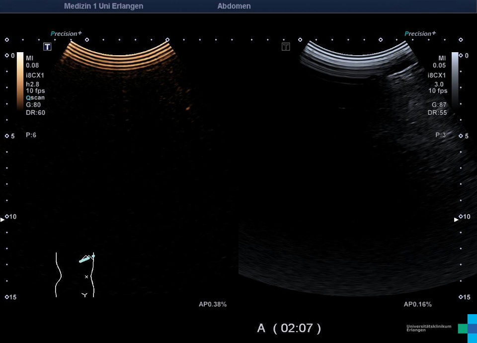
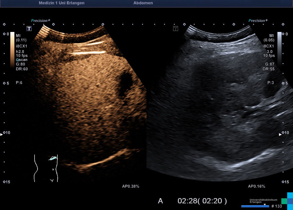
Epicrisis: status post Hepatitis C infection 10 years ago, noticeable hyperechoic lesions in ultrasound with normal vessels architecture -> typical liver lesions for porphyria cutanea tarda are hyperechoic and margin enhanced, which match focal steatosis. CEUS is used for ruling out a malignant lesion. The benignancy is proven by CEUS. Focal steatosis are isoenhanced in all CEUS phases. Clinical the patient shows no sign of skin lesions, but the patient reported such lesions after sun exposure. An association of HCV infection and porphyria cutanea tarda is reported in literature. With this patient the overall porphyrin in the 24h urine was elevated. Within the differentiation especially Uroporphyrin, Koproporphyrin, Hepttacarboxyporphyrin and Pentacarboxyporphyrin.
For better visualization due to didactic reasons the playback speed got reduced to 75%.


