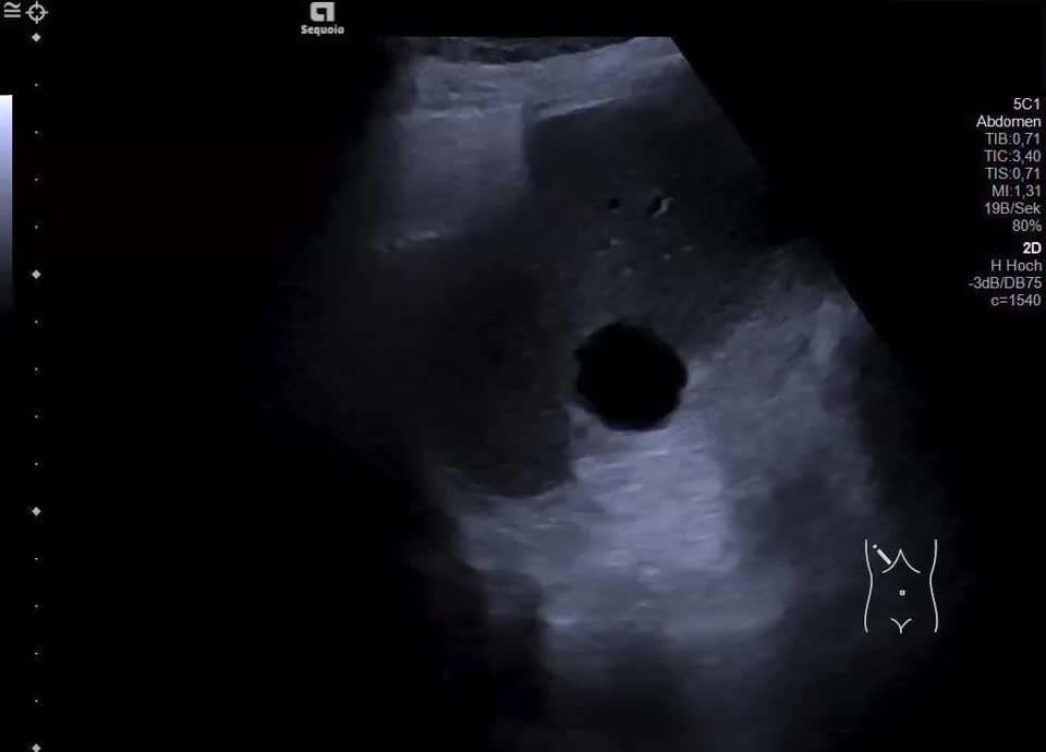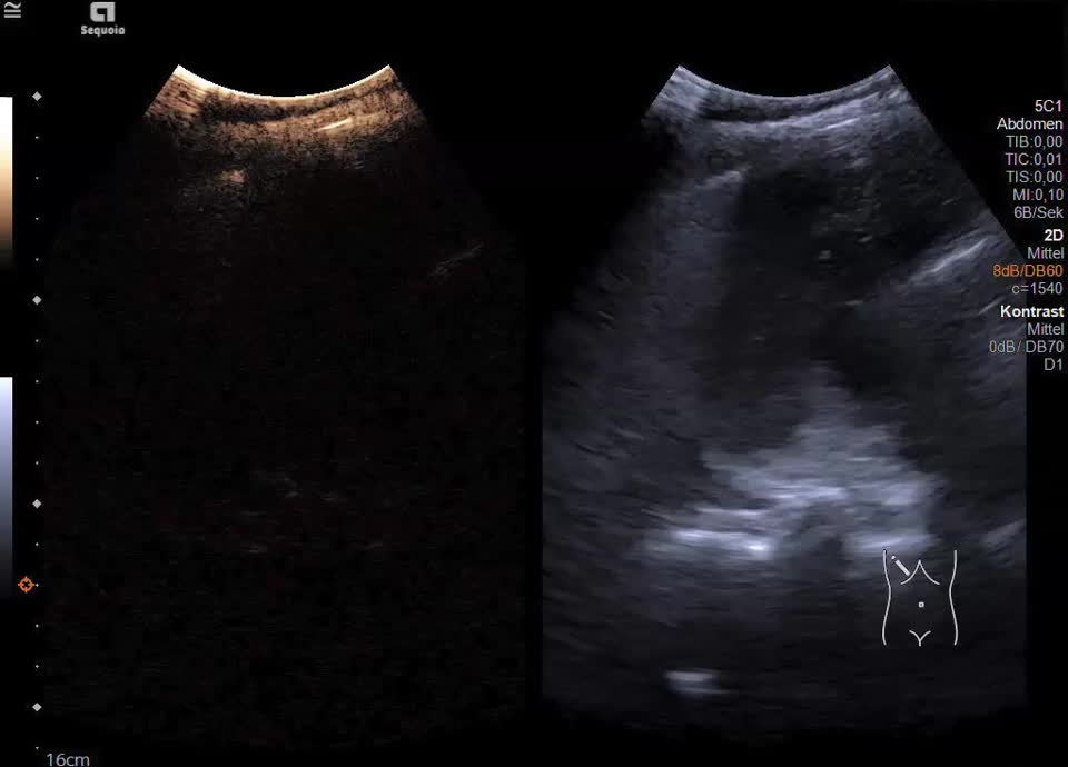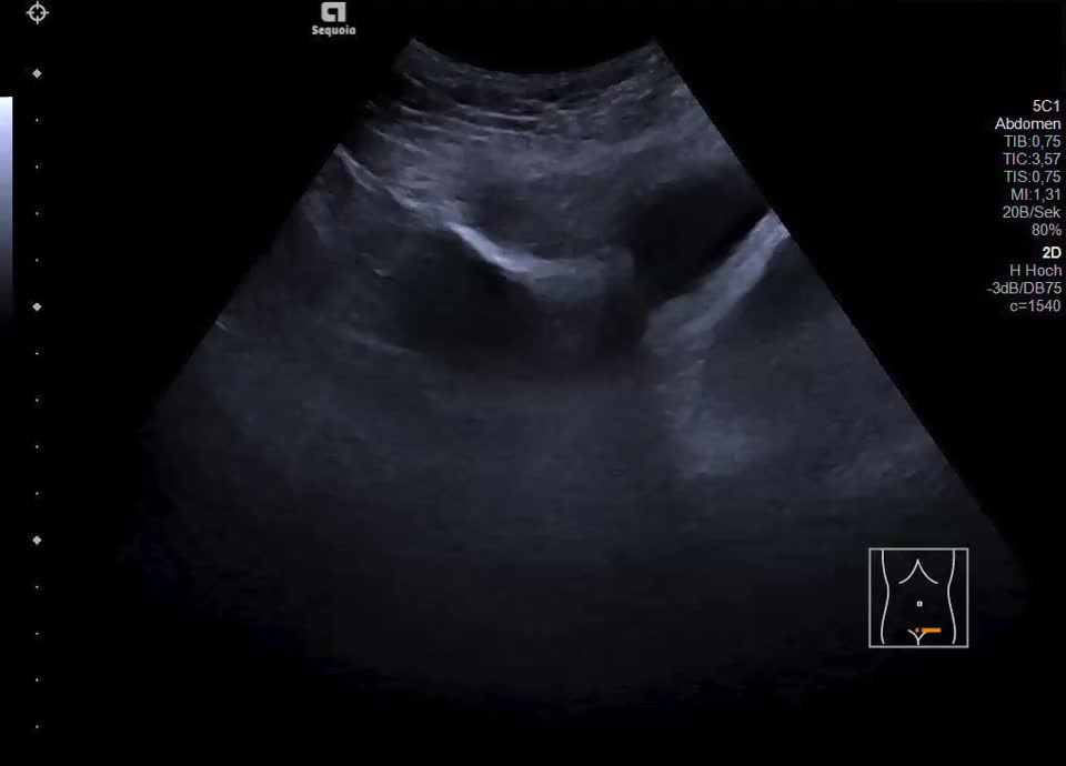

Cystic-solid liver metastasis in ovarian cancer
Case report: A 61-year-old female patient with suspected antiphospholipid syndrome, status post multiple cerebral infarctions and pulmonary embolism of unclear etiology, as well as a CT-morphologically indeterminate ovarian mass, underwent abdominal ultrasound evaluation. Ultrasound demonstrated a subcapsular cystic lesion in hepatic segment VI with a continuous capsular echo and an intralesional hyperechoic solid component. The findings are not consistent with a simple cyst but are suspicious for a cystic-solid metastasis. In the complementary CEUS, the solid components demonstrate contrast enhancement in the arterial phase and show persistent enhancement without washout in the portal-venous and late phases. Additionally, a nodular peritoneal thickening in the upper abdomen and a cystic ovarian tumor with solid components in the right lower abdomen are present. In summary, the findings are highly suggestive of a hepatic and peritoneal metastasizing ovarian carcinoma.