
Superinfected liver metastasis
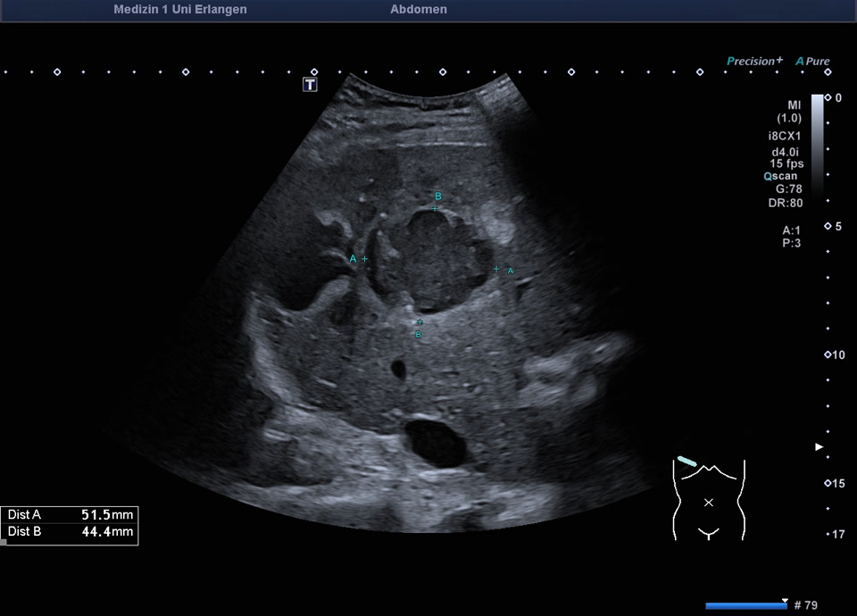
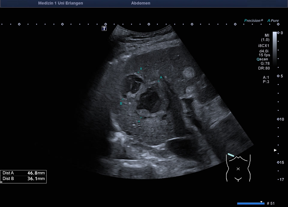
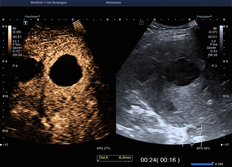
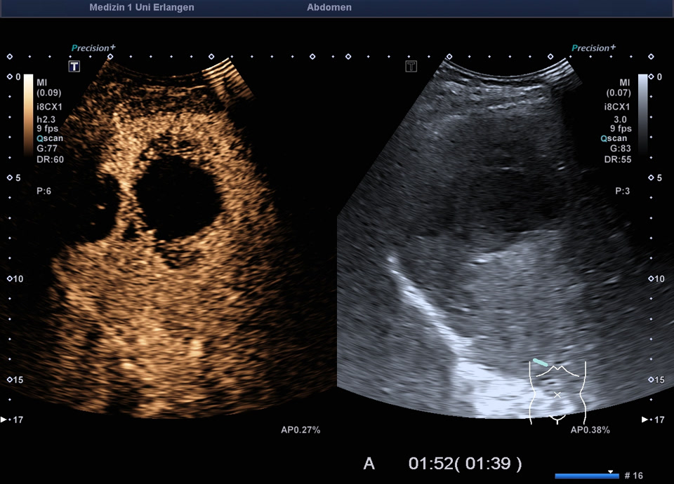
Case report: A 70-year-old female patient with known hepatic metastases from a histologically confirmed neuroendocrine tumor (NET G2) of the ileum, currently presenting with intermittent fever. An abdominal ultrasound was performed to rule out an abdominal source of infection. Ultrasound reveals the known multiple hyperechoic liver metastases in both hepatic lobes, as well as fluid-filled masses (previously described in the PET-CT from a year ago as metastases with central cavitation). On B-mode ultrasound, there is a strong suspicion of abscess formation with a transient arterial hyperemic rim on CEUS. In the portal venous phase, there is hypoenhancement of the surrounding liver parenchyma. The perfusion pattern, when considered in conjunction with the clinical presentation, raises suspicion of a liver abscess. During the subsequent drainage, turbid fluid containing old blood was drained; the working diagnosis is a hemorrhagic metastasis with secondary infection in the context of a known hepatically metastasizing NET.